ICMR BULLETIN VOL. 27-No.-10-OCTOBER-1997.pdf
Media
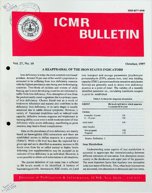
- extracted text
-
T
ISSN 0377-4910
ICMR
BULLETIN
Vol. 27, No. 10
October, 1997
A REAPPRAISAL OF THE IRON STATUS INDICATORS
Iron deficiency is today the most common nutritional
problem. Around 30 per cent of the world’s population is
estimated to be suffering from iron deficiency anaemia
with the highest prevalence rates being seen in developing
countries. Two-thirds of children and women of child
bearing age in most developing countries are estimated to
suffer from iron deficiency. Poor absorption of iron from
the predominantly cereal-vegetarian diet is a primary cause
of iron deficiency. Chronic blood loss as a result of
hookworm infestation and malaria also contribute to the
deficiency. Iron deficiency, in its early stages is usually
unnoticed due to subtle clinical symptoms. However, a
variety of functional disabilities such as reduced work
capacity, defective immune response and impairment in
learning ability occur even in mild-moderate states of iron
deficiency while severe deficiency, manifesting as gross
anaemia, may lead to foetal complications.
Data on the prevalence of iron deficiency are mainly
based on haemoglobin (Hb) estimations and there are
established norms to define anaemia in a population
(Table I). While a Hb level less than the expected for a
given age and sex is identified as anaemia, increase in Hb
levels even from the so called normal to higher levels
following iron supplementation may be accepted as an
indicator of iron deficiency in an individual. However, it
is not possible to obtain such information in all situations.
The precise definition of iron status that is reflected
in Hb levels needs to be identified. A battery of
haematological (Hb, haematocrit, RBC counts, etc.) and
iron transport and storage parameters [erythrocyte
protoporphyrin (EPP), plasma iron, total iron binding
capacity (TIBC), percent transferrin saturation and plasma
ferritin] are generally used to detect iron deficiency
anaemia at a point of time2. The validity of a recently
identified parameter viz., circulating transferrin receptor
is yet to be established.
Table I. Criteria for diagnosis of anaemia
GROUP
Hb levels (g/l) below which anaemia
is considered to exist
Children
6 months - 6 years
6-14 years
Adult males
110
120
130
Adult females
Non-pregnant
Pregnant
120
110
Source: Reference. 1
Iron Metabolism
Understanding some aspects of iron metabolism is
essential to appreciate the interrelationship between
different indicators of iron status. Iron absorption occurs
mainly in the duodenum and upper part of the jejunum.
The most important factor that regulates iron absorption
is the iron status of the body. Thus, when body iron reserves
are increased, iron absorption is decreased and vice versa.
In circulation iron binds to transferrin, its transport protein.
Transferrin bound iron through its cell surface receptor is
utilised by erythron for Hb synthesis. In erythroblasts,
iron is incorporated into pro.toporphyrin-IX ' for
haemoglobin synthesis and the rest of the body iron is
stored in ferritin.
Body Iron Compartments
The human body contains approximately 30-40 mg iron/
kg body wt. However, it varies as a function of age and sex
and physiological status. Of this, the storage iron
concentration ranges from 0 to 15 mg/kg body wt depending
on gender and iron status. The distribution ofthe stored iron
is not uniform and liver contains approximately 60 per cent
as ferritin. This storage iron has no known functions other
than acting as a reserve to replenish the lost iron from the
functional compartment. Iron in the functional compartment
is involved in cellular metabolism through incorporation
into haemoglobin, myoglobin, iron dependent enzymes and
other proteins involved in electron transport. About 85-90
per cent of the functional (non-storage) iron pool is in the
erythroid mass. The storage and functional compartments
are linked by a small transport compartment, through carrier
molecule transferrin.
Three stages of progression in iron deficiency have
been identified, using different iron status parameters and
the compartments affected2. The first stage is represented
by a depletion of the storage compartment as evidenced
either by a lowered marrow iron or levels of circulating
ferritin and is not reflected in any of the haematological
parameters-or those of iron metabolism (latent phase). The
second stage represents alterations in the transport system
in addition to depletion in stores and may be reflected in
decreased per cent transferrin saturation and increased EPP
(depleted erythropoietic pool). During the last stage, Hb
levels decrease and all parameters of iron status are af
fected (iron deficiency anaemia).
Limitations of Indicators
In all surveys to assess prevalence of iron deficiency,
Hb is used as the parameter which obviously indicates only
severe deficiency. Thus by clinical symptoms or routine
diagnostic parameters, the mildr-moderate degrees of iron
deficiency are missed. Identification and treatment of these
forms of iron deficiency is important to improve the qual
ity of life of a large population. All the haematological
and iron status parameters could singly identify severe
forms of deficiency. However, more than one measure
ment was suggested to identify mild-moderate forms of
iron deficiency, because the response of the parameters to
depletion was variable. Efforts to project circulating fer
ritin as an indicator of subclinical iron depletion were not
universally acceptable.
In a population, individuals are expected to be in a
state of change from either poor iron status to good status
or vice versa. Studies involving sequential monitoring of
various indicators including storage iron at different stages
ofdepletion and repletion along with bone marrow staining
for iron as a gold standard, are essential to understand the
relative merits and scope of each parameter. However,
such studies are not practical in human beings. The monkey
has been demonstrated to be a good model of ferrokinetics
and is phylogenetically closest to human beings. A series
of .studies were conducted in monkeys at the National
Institute ofNutrition (NIN), Hyderabad, to understand the
sequential changes in all iron status parameters during
different stages of depletion and repletion. In this article,
the results of these studies are highlighted and attempts
made to identify sensitive indicators of changes in iron
status.
Monkey Liver Ferritin
Monkey liver ferritin has been purified and
characterised for the first time and this was found to have
the close resemblance to human ferritin with regard to its
physicochemical properties. After raising suitable
polyclonal antibodies, an ELISA was developed for quan
tifying the serum ferritin as a measure of the iron status in
monkeys3.
Table II. Different modes of depletion and repletion of iron in
rhesus monkeys
Group
Mode
Duration
(months)
Depletion
Chronic
Short-term
Extended
Acute (phlebotomy)
Diet
Diet
Diet
Diet+blood loss
12
3
15
3
Iron-dextran injection
Diet
Diet
Diet
3
3
Repletion
Acute
Short-term (dietary adequate)
Short-term (dietary 50%)
Control
3
12
101
Indicators of Mild Iron Depletion
Mild iron depletion was produced in rhesus mon
keys (Macaco mulatto) fed a sejni synthetic iron deple
tion diet over a period of one year (Table II). Various
haematological and iron status parameters were moni
tored every month in all the animals. Liver iron was
estimated from the biopsy samples at the end of one
year. Bone marrow was aspirated at the same time
points and was subjected to iron staining.
Of the iron status indicators tested in milder forms of
iron deficiency (12 months), bone marrow iron staining,
haemoglobin concentration and haematocrit showed the
earliest change (Table III). Serum ferritin and liver iron
concentration did not respond to mild iron deficiency. Bone
marrow staining was very sensitive to depletion and indi
cated the highest correlation with changes in haemoglo
bin. A decrease in haematological parameters without
effecting any change in iron storage compartment during
mild depletion, indicates the existence of a “labile pool of
functional iron”, probably represented by erythroid iron.
This is more sensitive to changes in dietary iron and is in
equilibrium with bone marrow iron. Thus, the degree of
change in bone marrow grading is also reflected in hae
moglobin, haematocrit and RBC counts. It is concluded
from the study on rhesus monkeys fed on semi synthetic
iron depletion diet that early changes in haematological
parameters are adequate to identify mild forms of dietary
iron deficiency and parameters reflecting iron stores, in
cluding serum ferritin, are not sensitive to such a change4.
Table III. Changes in biochemical indicators of iron status in response to various modes of iron depletion and repletion in rhesus monkeys.
Group
Ht
Hb
Bone
marrow
iron
EPP
10m
11m
12m
9m
=
•=
Serum
iron
TIBC
% TS
12m
12m.
=
=
Serum
ferritin
Liver
iron
=
S3
Depletion
s
Chronic
Short-term
=
=
=
90d
==
Extended
15m
14m
15m
30d
30d
90d
30d
60d
30d
+
90d
60d
Short-term
(dietary
adequate)
+
30d
30d
+
90d
30d
Short-term
(dietary 50%)
+
30d
30d
90d
30d
Acute
(phlebotomy)
=
30d
=
=
=
=
14m
14m
+
60d
30d
+
30d
=
30d
s
=
=
=
=
=
Repletion
Acute
=
=
60d
=
=
Ht: Haematocrit; Hb: Haemoglobin; EPP: Erythrocyte protoporphyrin; TIBC: Total iron binding capacity;
%TS: Per cent transferrin saturation
m:
Months; d : Days when first changes were seen.
- Decrease; + Increase; = No change
102
90d
This is contrary to the classical theory that the first and
second stages of deficiency are not reflected in Hb status.
Different Modes of Depletion and Repletion
Mildly anaemic monkeys were subsequently continued
on the iron depletion diet or were repleted with different
supplements of iron for 3 months. Control animals were
placed on an iron deficient diet or further subjected to
periodic phlebotomy (Table II).
Effect of Iron Depletion
In the early stages of depletion, only bone marrow iron
was found to be decreased even before a decrease in
haemoglobin (Tablelll). This finding supports the classical
definition of latent phase of deficiency to the extent that a
decrease in marrow iron, is the first change during
depletion. Chronic and extended dietary depletion resulted
in decreased haemoglobin along with bone marrow iron
(Table III). Decrease in serum iron and percent saturation
were also noted in such dietary depletion. On the other
hand, all haematological as well as iron status parameters
responded to acute depletion involving phlebotomy: None
ofthese manipulations for iron depletion disturbed either
serum ferritin concentrations or liver iron concentration,
representing iron stores.
Effect of Iron Repletion
During various modes of repletion, increase in haemo
globin was the earliest sign along with that of marrow iron
followed by other changes like decrease in EPP and increase
in transferrin saturation. Liver iron increased only in acute
repletion by intramuscular injection of iron and moderate
supplements through diet did not result in any change in liver
iron. Serum ferritin and liver iron did not respond to supple
ments with dietary iron. Only repletion through iron-dextran
injection had a positive effect on all the haematological and
iron transport indicators. Under this condition liver iron also
responded but not serum ferritin. Thus, different parameters
responded variably under different modes of depletion/repletion, as their response is perhaps related to the degree and
duration of change in iron nutriture that had been taking place
before and during the intervention5.
Significance of Iron Status Parameters
The turnover of iron in a given compartment* may
depend on the pre-existing iron status of the animal and
subsequent response of the indicators may depend on the
immediate demands, pool size of the compartments and
the regulatory mechanisms operating at that time.
Haemoglobin stands out as the first and best
haematological parameter to reflect the changes in iron
status as corroborated by bone marrow iron. This is
obvious when two-thirds of body iron is present in
haemoglobin and its turnover is much greater than that of
stored iron.
While the three stages of iron deficiency suggested
earlier provide a convenient conceptual framework of
dynamics of iron depletion, evaluation of prevalence
data for anemia contrpl programmes in humans did not
conform strictly to this pattern in individuals with mild
iron deficiency. A decrease in serum ferritin along
with bone marrow iron even before a decrease occurred
in haemoglobin has been shown as evidence for exist
ence of a “latent” stage of iron deficiency. Only in
phlebotomy models of iron depletion was such sensi
tivity of ferritin demonstrated by other investigators.
In a representative sub-sample comprising 200 subjects
in a field study carried out in about 6000 rural
Hyderabad population, a direct relationship was seen
only between cases of hypoferritenaemia (<12 ng/ml)
and severe forms of anaemia (Hb <1 Og/dl; NIN, un
published data). But no direct proof was available re
lating serum ferritin to liver or bone marrow iron in
conditions where dietary depletion was present. Most
of the sequential studies in rats also did not support such
a latent phase6. The insensitivity of serum ferritin to
phlebotomy in the present studies could be explained
on the basis of unaltered liver iron stores observed in
short-term depletion and repletion.
Dynamics of Iron Balance and Compartments
Iron deficiency may develop suddenly or gradually.
Acute depletion, as with phlebotomy, leads to a rapid
exhaustion of stored iron (through increased rates of
haemoglobin synthesis) and this may lead to redistribution
of the remaining body iron. Chronic depletion is a gradual
process and normally occurs due to inadequate amounts
of available iron in the diet and may induce iron deficiency
with simultaneous depletion of all iron stores.
A revised model incorporating a labile pool to explain
the observations noted here is proposed in the figure. The
upper panel provides a schematic representation of move
ment of iron among the different compartments during
103
SONt
AUHOW
LABILE
POOL
i
eroiu&t
r—•
1 a "
gKYTHKQlD VKANSPOHf
POOL *
PL Atm
»
Fig. Schematic representation of iron compartments (upper panel)
and stages operating during depletion or repletion (lower
panel).
The scheme indicates change of L followed by B before any change
in S during depletion or repletion.
(B. Bone marrow, L. Labile pool, S. Storage, A. Absorption)
1.
Normal with good stores
2.
Depletion of labile pool before any change in stores
3.
Depletion of storage pool
4.
Repletion of labile pool without any change in stores
depletion and repletion. The central concept is that the
“labile pool” of iron is the first pool to get depleted or
repleted before the stores are changed. For this to occur,
movement of iron during repletion is not just opposite to
that in depletion and the new scheme (lower panel)
accomodates the observed sequential changes in different
iron pools. Stage 1 represents the normal iron status
wherein all the iron compartments are in equilibrium and
are operating at the optimum level. In the course of deple
tion, stage 2 indicates exhaustion of the labile pool (L)
which is considered to be in immediate equilibrium with
haematopoietic bone marrow pool (B), before any change
occurs in storage pool (S). Exhaustion of the labile pool
triggers mobilization of stored iron from the liver and
spleen as depicted in stage 3. The cut-off in the supply of
104
labile pool restricts channeling of iron into bone marrow
dependent functional activity ie., decrease in haemoglo
bin synthesis and RBC formation and explains the delay
in accumulation of EPP. On the other hand, repletion fills
the labile pool first and then promotes haemoglobin syn
thesis simultaneous with lowering of EPP as in stage 4.
Ultimately the stores are also repleted on continued supple
mentation reaching a normal state (stage 1). The present
studies provide support for the model suggested by
Pollycove and Mortimer7 that ferrokinetics follow a two
compartmental model during depletion and three com
partmental model during repletion
It is conceivable that if a'person is found to have 10 g/
dl haemoglobin at a given time, one of the following three
situations could arise with regard to the background of the
subject: (i) The subject has been stabilised with mild iron
deficiency anaemia over a period of time; (ii) the subject
had a good iron status earlier but due to the change in the
intake, is on a course of depletion at the time of observa
tion; or (iii) the subject, to start with, had iron deficiency
anaemia but, is in the process of recovery at the given
moment.
Monitoring the iron status (at least at two points) could
normally be expected to provide such information. How
ever, actual data did not provide any such clues nor was
any analysis really attempted.
As indicated from these experiments, if it is a fact
that various iron status parameters do respond
differently during repletion and depletion, it is
possible theoretically to use more than one parameter
along with haemoglobin to obtain information on
the previous iron status of the subject in relation to
the current condition. Viewed in this context, EPP
seems to be sensitive to repletion rather than
depletion. Similarly transferrin saturation seems to
respond to depletion better than repletion. These
results may be reflecting the typical sensitivity of
the indicators and the changes taking place in the
iron compartments. The present studies highlight the
potential of obtaining wholesome information on iron
status using selected parameters. With more vigorous
testing it is possible to identify the ideal combination
of tests which will yield a comprehensive picture of
the iron status of an individual with a single sample
at one time point.
References
1.
WHO. Nutritional anaemias. WHO Tech Rep Ser 503: 29,1972.
2.
Interpretation of iron status measurements. In: Measurements
ofIron Status. A Report of the International Nutrition Anaemia
Consultative Group (1NACG), Nutrition Foundation Inc, Wash
ington, DC, p.72, 1985.
5.
Sreeramulu, D., Madhavan Nair, K., Qadri, S.S.Y.H. Visweswara
Rao, K. and Sivakumar, B. Changes in biochemical indicators
of iron status during iron repletion and depletion in monkeys.
Ann Nutr Metab 41: 126, 1997.
6.
Hunter, J.E. Variable effects of iron status on the concentration
of ferritin in rat plasma, liver and spleen. J Nutr 108: 497,
1978.
Pollycove, M. and Mortimer, R. I he quantitative determina
tion of iron kinetics and haemoglobin synthesis in human sub
jects. J Clin Invest 40: 753, 1961.
3.
Sreeramulu, D., Madhavan Nair, K. and Sivakumar, B. Isolation
and characterization of monkey liver ferritin. Indian J Biochem
Biophys 33: 48, 1996.
7.
4.
Sreeramulu, D., Qadri, S.S.Y.H., Madhavan Nair, K. and
Sivakumar, B. Changes in serum ferritin and other biochemical
indicators of iron status. Ann Nutr Metab 38: 322, 1994.
This write-up has been contributed by Dr. B. Sivakumar, Deputy
Director (Sr.Grade) and K. Madhavan Nair, Senior Research
Officer, National Institute of Nutrition, Hyderabad
ABSTRACTS
Some Research Projects Completed Recently
Molecular fingerprinting of bacteria causing
nosocomial infections.
Plasmid fingerprinting was applied for tracing the
source of nosocomial infection with plasmid containing
bacteria. In addition, bacterial chromosomal fingerprint
ing after, restriction endonuclease digestion was done to
determine the relationship between isolates of bacteria
causing nosocomial infection but not containing plasmids.
Forty seven strains of Klebsiella pneumoniae were
subjected to klebocin typing by scrape and streak method
using 6 indicator strains. All the strains were found to be
positive to klebocin thereby offering no discrimination.
By klebocin extraction method these 47 strains could be
grouped into 11 distinct klebocin types. However, on
repeated testing only 35 strains gave similar pattern
indicating a reproducibility of 74.5 per cent. Three major
categories of klebocin types predominated even in
epidemiologically unrelated strains making discrimination
difficult. The two outbreaks (August 1994, June 1995)
investigated were caused by a klebocin type designated
444, a group indicating resistance to klebocin produced
by all six producer strains and essentially untypable by
klebocin typing. Whole cell protein analysis (WCPA) by
SDS-PAGE could subdivide the 7 strains from the first
outbreak into 5 groups while the 3 strains from the second
outbreak were found to be homogenous. Ribotyping of the
same isolates indicated 5 groups in the first outbreak and
2 groups in the second outbreak were found to be
homogenous. Ribotyping of the same isolates indicated
5 groups in the first outbreak and 2 groups in the second.
Ribotyping had better discrimination and indicated that
these outbreaks were actually pseudo-outbreaks and notw
common source outbreaks as was implied by the use of
klebocin typing.
Forty two strains ofPseudomonas aeruginosa isolated
from bronchoalveolar lavage from intubated patients
between December 1993 to June 1994 were also analyzed.’
Pyocin typing of these strains by scrape and streak method
using 22 indicator strains revealed excellent discrimination
in 40 strains. But on repeated testing the group designation
changed indicating that the system had low reproducibility.
SDS-PAGE of whole cell protein profile indicated presence
of 45 protein bands of different molecular weights,
individual isolates had 37 to 42 protein bands ranging from
340 to 143 kDa. On the basis of Dice Index of similarity
the strains could be grouped into 20 types. Since all strains
could be typed the system had adequate typability. Similar
results were obtained on repeated testing indicating good^J:
reproducibility. Ribotying of these isolates could group
them into 18 groups; the major group comprised 8 isolates.
It is thus clear, that pyocin typing had excellent dis
crimination but poor to no reproducibility while klebocin
typing had adequate reproducibility but not sufficient dis
crimination. Therefore, these methods cannot be of use
for reliable fingerprinting of isolates leading to tracing of
the source of infection in an outbreak. Whole cell protein
analysis by SDS-PAGE offered a reliable, reproducible
and fairly discriminating method for identification of
clones of bacteria.
Plasmid analysis was not found to be a very
discriminatory or sensitive indicator for fingerprinting. But
105
ribotyping, using a DIG labelled 18 mer oligonucleotide
probe complementary to a conserved region of the
16 S rRN A gene showed good results. The probe was stable
and hybridized with DNA extracted, from a wide variety
of bacteria. Since the rm operon is present in multiple
copies in the DNA of each bacteria, a number of bands
would light up making determination of the clonal nature
of the isolates easy. Discrimination could be improved by
selection of more than one restriction endonuclease for
cutting the bacterial DNA.
i
A. Rattan
Department of Microbiology
All India Institute of
Medical Sciences
New D^lhi.
^Publications:
1.
Khan, I.A., Rattan, A., Khan, F.G. and Kaiia, A. Fingerprinting
ofSalmonella typhimurium - comparison between traditional and
molecular methods. Indian J Med Microbiol 13: 141, 1995.
2.
Khan, I.A., Rattan, A., Fatima, T., Khan, F.G. and Kaiia, A.
Application of whole cell protein analysis by SDS-PAGE to
establish the source of Salmonella typhimurium. J Infect 33: 169,
1996.
3.
Khan, F.G., Rattan, A., Khan, I.A. and Kaiia, A. A preliminary
study of fingerprinting of Pseudomonas aeruginosa by whole
cell protein analysis by SDS-PAGE. Indian J Med Res 104:342,
1996.
Immunological studies in patients with cervical lesions
using recombinant human papillomavirus fusion proteins.
21 patients of CIN-II, 13 patients of CIN-III, 30 patients of
histopathologically confirmed carcinoma cervix and 30
healthy asymptomatic women. Two synthetic peptides of
HPV16E7 comprising the amino acid sequences
QAEPDRAHYN1VTF (a,a. 44-57) and QLNDSSEEEDEI
(a.a. 27-38) were designated as peptides PI and PI1
respectively. Thirteen (43.3%), 4(19%), 7(53.8%) and
8(26.6%) patients with CIN-I, CIN-II, CIN-III and carcinoma
cervix respectively had antibodies to Pl peptide whereas
9(30%), 16(76%), 6 (46.1 %) and 18(60%) patients with CINI, CIN-II, CIN-III and carcinoma cervix respectively had
antibodies to PI I peptides. None of the healthy women had
antibodies to PI and PII peptides of HPV16E7.
Lymphoproliferative assays (LPA) were carried out with
synthetic peptide (EPDRAHYNIVTFCCj a.a. 46-59) of
HPV16E7 T cell epitope for detection of T cell responses.
Two (6.6%) of the 30 controls and 19 (63.3%), 9( 42.8%),
4(30.7%) and 11(36.6%) patientswith CIN-I, CIN-II, CINIII and carcinoma cervix respectively showed positive
response to T cell epitope. No correlation could be established
between the severity of the disease and T cell responses.
The study thus suggests that peptides PI and PII are the
two major immunodominant B cell epitopes ofthe HPV 16E7
and PII is more immunodominant compared to PI. However,
more studies need to be carried out to see whether antibody
detection to these two peptides can be used to monitor the
prognosis and biological progression of patients with CIN.
Serum samples from 13 histopathologically confirmed
patients with carcinoma cervix and 9 healthy asymptom
atic women were analysed for detection of antibody re
sponse to HPV16E7 and LI fusion proteins in a Western
blot assay. Antibodies to HPV16E7 fusion protein were
detected in 9 (69.2%) patients and 1 (11.1%) healthy
woman, whereas all the 13 patients and 9 healthy women
revealed the presence of antibodies to HPV16L1 fusion
protein.
DNA samples from biopsy material from 30 carcinoma
cervix patients were tested for the presence of HPV DNA in
dot blot hybridization experiments. Twenty three (76.6%)
DNA samples were positive for HPV DNA under low strin
gency conditions and 10(43.4%) ofthese 23 samples showed
the presence of HPV 16 DNA. ELISA showed the presence
of antibodies to HPV 16E7 in 7 (70%) of the 10 patients with
HPV16DNA, 6 (46.1%) of the 13 patients with HPV DNA
and 6 (85.7) of the 7 patients with no evidence of HPV DNA
by dot blot hybridization. LPA reactivity to HPV 16E7 T cell
epitope was observed in 3 of 10 (30%) HPV 16 positive, 7 of
13 (53.8%) HPV 16 negative but other HPV positive and 1 of
7 (14.3%) HPV negative cervical cancer patients. These find
ings suggest that LPA reactivity to HPV16E7 T cell epitope
was depressed in most HPV 16 positive cervical cancer pa
tients.
ELISA was carried out with the synthetic peptides of
two B cell epitopes of HPV16E7 gene product for detecting
the presence of antibodies in 30 patients cytopathologically
confirmed to have cervical intraepithelial neoplasia (CIN)-I,
P. Seth
Department of Microbiology
All India Institute of Medical Sciences
New Delhi.
The study was carried out to determine the humoral and
cellular immune response against human papillomavirus
(HPV) 16 in patients with benign and malignant lesions of
| the uterine cervix and to correlate the response with the clini
cal status of the patients.
106
ICMR NEWS
The following meetings of various technical coipmittees/groups of the Council were held:
the XVIII International Vitamin A Consultative Group
meeting at Cairo (September 22-26, 1997).
Meetings of the Task Force (TF)ZProject Review
Committee (PRC) held at New Delhi:
Dr. J.M. Deshpande, Director, Enterovirus Research
Centre, Mumbai, participated in the meeting of Global
Polio Laboratory Network at Geneva (September 25-26,
1997).
TF on Case Control Study to
Identify Risk Factors for
Acute Myocardial Infarction
in Indians
September 30October 1, 1997
PRC on Oncology and Pathology
October?, 1997
Participation of ICMR Scientists in Scientific Events:
Dr. Manjula Datta, Dy. Director, TRC, Chennai, par
ticipated in the meeting of the Working Group for the
Development of New TB Vaccine of the WHO Global
Programme for Vaccines and Immunization/Vaccine Re
search and Development at Geneva (October 4, 1997).
Dr. V.K. Vijayan, Dy. Director (Sr. Grade), TRC,
Chennai and Dr. A.K. Mukherjee, Sr. Research Officer,
Regional Occupational Health Centre, Calcutta, partici
pated in the IX International Conference on Occupational
Respiratory Diseases at Kyoto (October 13-16, 1997).
Dr. V. Kumaraswami, Dy. Director, Tuberculosis
Research Centre (TRC), Chennai, participated in the
II meeting of the Task Force on Community Directed
Treatment of Lymphatic Filariasis and Onchocerciasis at
Ouagadougou, Burkino Faso (September 16-19,1997).
Honours/Awards:
Dr. P. Bhaskaram and Dr. B. Sivakumar, Dy. Directors
(Sr.Grade) and Dr. K.V.R. Sarma, Asstt. Director, National
Institute of Nutrition, (NIN), Hyderabad, participated .in
Dr. Kamala Krishnaswamy, Director, NIN, Hyderabad,
has been elected as Fellow of the Indian National Science
Academy, New Delhi.
ICMR AIDED SYMPOSIA/SEMINARS/WORKSHOPS/COURSES/CONFERENCES
Symposium/Seminar/WorkshopZ
Course/Conference
Date & Place
Contact Address
IV National Conference of the Society of Bio
materials and Artificial Organs on New Horizons
in Biomaterials Research
October 3-4, 1997;
(at Hyderabad)
Dr. P.R. Chatteijee, Convenor of the Conference,
Indian Institute of Chemical Technology, Hydera
bad-500007.
IX National Congress of Veterinary Parasitology
October M, 1997;
(at Ludhiana)
Dr. H.S. Bali, Organising Secretary of the Cong
ress, Department of Veterinary Parasitology, Col
lege of Veterinary Science, Punjab Agricultural
University, Ludhiana-141004.
Guha Research Conference-1997
October 13-17, 1997;
(at Palampur)
Dr. Amit Ghosh, Co-convenor-GRC’97, Institute
of Microbial Technology, Chandigarh-160014.
Seminar on Current Status of Thalassaemia in
India and National Workshop on Carrier
Detection and Antenatal Diagnosis of
Thalassaemia
November 13 and
14-15, 1997;
(at Lucknow)
Dr. Sarita Agarwal, Organising Secretary, Dep
artment of Medical Genetics, Sanjay Gandhi
Postgraduate Institute of Medical Sciences, Lucknow-226014.
International Conference on Advances in
Reproductive Medicine
November 15-18,1997;
(at Calcutta)
Dr. Sudersan Ghosh Dastidar, Organising Secretary-ICARM’97, Infertility Clinic and I.V.F. Cen
tre, 208, Rash Behari Avenue, Calcutta-700029.
Asia-Pacific Congress on Head and Neck Cancer
December 11-14,1997;
(at Mumbai)
Dr. Ashok R. Mehta, Chairman of the Congress,
Department of Surgery, Tata Memorial Hospital,
Mumbai-400012.
-in-7
COUNCIL’S TRAINING PROGRAMMES FOR 1997-98
Reproductive Biology
•
At the Institutefor Research in Reproduction, Mumbai:
• Workshop on Basic Molecular Biology for Clinicians
(December 2-11, 1997).
Training Course on Environmental Hygiene Practice
(November 10-14, 1997).
Laboratory Animal Technology
Nutrition
At the National Centre for Laboratory Animal Science,
National Institute ofNutrition, Hyderabad:
At the National Institute of Nutrition, Hyderabad:
•
•
Postgraduate Certificate Course in Nutrition (Decem
ber 1, 1997-February 28, 1998).
Training Course for Laboratory Animal Supervisors
(September 1 -November 29, 1997).
Haematology
Occupational Health
At the Institute of Immunohaematology, Mumbai:
At the National Institute of Occupational Health,
Ahmedabad:
•
Training Course in Transfusion Medicine for Blood
Bank Medical Officers (August 27-October 24,
1997).
•
Training Course in Advanced Haematology and
Immunohaematology (October 6-24,1997).
•
Orientation Course on Occupational Health for Indus
trial Medical Officers (September 30-October 11,
1997).
ICMR PUBLICATIONS
Nutritive Value of Indian Foods (1985), by C. Gopalan, B.V. Ramasastri and
S.C. Balasubramaniam, Revised and Updated (1989), by B.S. Narasinga Rao, K.C. Pant
and Y.G. Deosthale (Reprinted 1995)
Price (Rs.)
23.00
Growth & Physical Development of Indian Infants and Children (1972, Reprinted 1989)
10.00
Studies on Weaning & Supplementary Foods (1974, Reprinted 1996)
15.00
Studies on Pre-School Children (1974, Reprinted 1984)
6.00
A Manual of Nutrition (Second Edition 1974, Reprinted 1995)
6.00
Low Cost Nutritious Supplements (Second Edition 1975, Reprinted 1994)
4.00
Menus for Low Cost Balanced Diets and School Lunch Programmes Suitable for
North India (Second Edition 1977, Reprinted 1994)
4.50
Menus for Low Cost Balanced Diets and School Lunch programmes Suitable for
South India (Fourth Edition 1996)
6.00
Some Common Indian Recipes and their Nutritive Value (Fourth Edition 1977,
Reprinted 1995) by Swaran Pasricha & L.M. Rebello
10.00
Nutrition for Mother & Child (Third Edition 1978, Reprinted 1994) by P.S.
Venkatachalam & L.M. Rebello
9.00
Some Therapeutic Diets (Fifth Edition 1996) by Swaran Pasricha
7.00
Nutrient Requirements & Recommended Dietary Allowances for Indians (1990, Reprinted 1995)
16.00
Fruits (Second Edition 1996) by Indira Gopalan & M. Mohan Ram
20.00
108
Count What You Eat (1989, Reprinted 1994) by Swaran Pasricha
9.00
Diet & Diabetes (Second Edition 1993) by T.C. Raghuram, Swaran Pasricha & R.D. Shanna
18.00
Dietary Tips for the Elderly (1992) by Swaran Pasricha & B.V.S. Thimmayamma
3.50
Diet and Heart Disease (1994, Reprinted 1996) by Ghafoorunissa and Kamala Krishnaswamy
26.00
Depressive Disease (1986) by A. Venkoba Rao
58.00
•Medicinal Plants of India Vol.2 (1987)
136.00
* 25 per cent discount allowed to individuals.
These publications are available on prepayment of cost by cheque, bank draft or postal order (bank and postal charges will be
extra) in favour of the Director-General, Indian Council of Medical Research, New Delhi. Money orders are not acceptable. All
correspondence in this regard should be addressed to the Chief, Division of Publication and Information, Indian Council of Medical
Research, Post Box No 4911, Ansari Nagar, New Delhi-110029 (India).
EMERITUS MEDICAL SCIENTISTS
INDIAN COUNCIL OF MEDICAL RESEARCH
Nominations/applications are invited from distinguished retiring scientists engaged in research
in the field of biomedicine for consideration for appointment as Emeritus Medical Scientists under the
Council. Full particulars can be obtained from the office of the Director-General (Personnel Section),
Indian Council of Medical Research, Post Box No. 4911, New Delhi-110 029. Scientists who have
retired or are going to retire by 31st March, 1998 may apply for the appointment as Emeritus Medical
Scientist under the Council.
Last date for receipt of applications is 30th November, 1997.
EDITORIAL BOARD
Members
Chairman
Dr. Badri N. Saxena
Dr. Bela Shah
Dr. Lal it Kant
Director-General
ICMR
Editor
Dr. N. Medappa
Printed and Published by Shri J.N. Mathur for the Indian Council of Medical Research, New Delhi
at the ICMR Offset Press, New Delhi-110 029
R.N. 21813/71
Position: 2419 (10 views)