ICMR BULLETIN VOL.5-No.-11-NOVEMBER-1975
Item
- Title
- ICMR BULLETIN VOL.5-No.-11-NOVEMBER-1975
- extracted text
-
health cell
326, V Main, I Block
Koramtrngala
Bangalore—560034
India
Vol. 5 No. 11
ANAEMIA
IN
Anaemia in pregnancy is a major public
health problem in developing countries. While
anaemia is directly responsible for 20% of
maternal deaths,in another 20% it is a contri
buting factor (Menon,M.K.K.,1967). Besides,it
is also responsible for the high incidence of
premature and low weight births thus increas
ing the perinatal mortality and morbidity.
Studies carried out at the National Institute
of Nutrition, Hyderabad are highlighted below:
HAEMOGLOBIN SURVEYS:
Haemoglobin
surveys
carried
out in
different parts of the country indicate that
while 15—20% of women are anaemic at the onset
of pregnancy, the incidence of anaemia (ilb.
levels less than 11 g%) increases to 60-70% in
the last trimester.
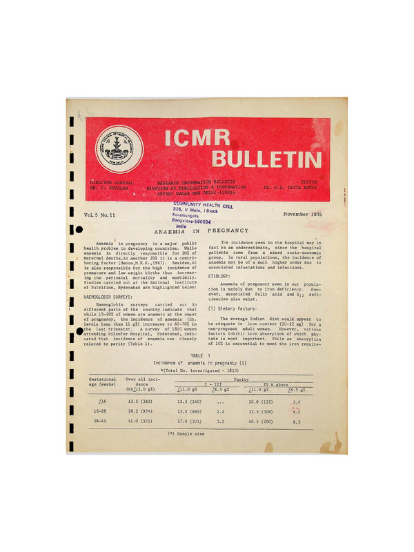
A survey of 1810 women
attending Niloufer Hospital, Hyderabad, indi
cated that incidence of anaemia was closely
related to parity (Table 1).
TABLE
Incidence of
November 1975
PREGNANCY
The incidence seen in the hospital may in
fact be an underestimate, since the hospital
patients come from a mixed socio-economic
group. In rural populations, the incidence of
anaemia may be of a much higher order due to
associated infestations and infections.
ETIOLOGY:
Anaemia of pregnancy seen in our popula
tion is mainly due to iron deficiency.
How
ever, associated folic acid and Bj2 deficiencies also exist.
(1) Dietary factors:
The average Indian diet would appear to
be adequate in iron content (20-22 mg) for a
non^pregnant adult woman.
However, various
factors inhibit iron absorption of which phy
tate is most important. While an absorption
of 10% is sessential to meet the iron require
1
anaemia in pregnancy (%)
*(Total No. investigated - 1810)
Gestational
age (weeks)
Over all inci
dence
(Hb_/11.0 g%)
/11.0 g%
/16
15.5 (265)
12.5 (140)
16-28
29.2 (974)
23.0 (666)
28-40
41.0 (571)
37.0 (371)
Parity
(*) Sample size
I -Ill
Z8.5 g%
IV & above
/_11.0 g%
£8.5 g%
20.0 (125)
,2.0
2.2
32.5 (308)
• "6:0
3.2
48.5 (200)
8.5
ment of a normal adult, studies using whole
body counter indicate that only 3-5% of die
tary iron is absorbed in an apparently normal
healthy individual. The diet is deficient in
protein, calcium and vitamin C. The diet is
also deficient in other haemopoietic factors
like folic acid and B】2・
The iron needed during pregnancy is of
the order of 4-6 mgs of absorbed iron daily,
which cannot be made available through dietary
sources. Since most women do not receive any
supplements during pregnancy, iron deficiency
is a common feature.
(2) Iron stores and Iron Ioss:
Most of the population in Asian Countries
have very poor iron stores as indicated by
poor bone marrow haemosiderin and low levels
of liver iron.
Besides, iron losses through
menstruation, to the extent of 15-30 mg every
cycle, increases the iron requirement of adult
women. Most women enter their first pregnancy
with very little iron reserve.
(3) Demands of Pregnancy:
-The net iron loss during pregnancy in
Indian women is around 350 mg.
To meet this
additional demand, a woman needs to absorb
2-3 mg of additional iron daily. Besides the
increased demands during pregnancy, demands
during lactation contribute to reduce iron
stores and worsen the deficiency status. Since
many women have repeated, closely spaced preg
nancies with prolonged periods of lactation,
there is progressive depletion of iron stores
with increasing numb er of pregnancies.
This
results in high incidence of anaemia in women
with higher parities.
(4) Infections and Infestations:
f
Repeated and chronic infections prevent
the utilization of iron and are additional
contributory factors in causing anaemia. Hook
worm infection in some areas aggravates the
situation by increasing iron loss.
ABSORPTION STUDIES:
Iron absorption:
Studies carried out on food iron absorp
tion at the National Institute of Nutrition
have indicated that during pregnancy, iron
absorption from food increased from an average
of 10% in the first trimester to 25-30% at
term. In addition, iron absorption was of a
2
much greater order in women in whom the trans
ferrin saturation was less than 15%. On the
basis of these data, iron requirement during
pregnancy has been computed to be around 40 mg
per day. However, the requirement is greater
in women who have very poor iron stores and
]ow haemoglob in levels at the onset of preg
nancy .
Folic acid absorption:
There is considerable contraversy regard
ing absorption of folic acid in pregnancy. It
is believed that folic acid deficiency seen in
pregnancy is mainly due to impaired folic acid
absorption and partly due to increased renal
clearance of
folate.
Transfer of folate
across the placenta to the foetus is also con
sidered as an additional cause.
Studies carried out on folic acid absorp
tion using tritiated folic acid in women
undergoing therapeutic termination of preg
nancy in the first and second trimester, at
the National Institute of Nutrition have indi
cated that there is no impairment in the
absorption of crystalline folate during the
first and second trimesters of pregnancy. How
ever, whether this is true of food folate
needs to be investigated. These studies also
suggest that the folate status of the indivi
dual influenced the extent of absorption but
this observation needs confirmation.
Serum levels of iron, folic acid and B12:
in spite of normal haemoglobin levels,
about 50% of pregnant women have low levels of
serum iron, it being less than 60 ug/100 ml in
the last trimester. The percentage transferrin
saturation is also low in these women.
Determination of serum folic acid and Bj2
levels have indicated that in about 60% of
women, serum folate levels were less than
3 ng/ml indicating folate deficiency and that
in 25% levels of serum B】2 were below 100 ng/
ml in the last trimester.
In a small number of women with severe
anaemia, where haemoglobin levels were less
than 8.5 gms%, studies on bone marrow indi
cated that in 60% of them there was megablastic erythropoiesis.
Folate levels in RBC in
these women was below 80 ng/ml. There was no
stainable iron in the bone marrow in any of
the subjects.
These observations suggest that while in
mild and moderate degrees of anaemia in preg
nancy iron deficiency was the major factor, in
the severe forms folate deficiency is clearly
present.
TREATMENT AND PREVENTION OF ANAEMIA:
EFFECT OF MATERNAL ANAEMIA ON FOETUS
BORN:
In cases of severe anaemia, during the
last trimester of pregnancy, there is very
little time to improve the haemoglobin and
iron status with oral or parenteral iron. In
addition, a greater majority of such women
come in a state of anoxia and cardiac failure.
In such cases, an
initial transfusion of
packed cells or exchange transfusion needs to
be given, followed by supportive treatment to
improve her nutritional status. With the ad
vent of packed cells or exchange transfusion,
maternal mortality rate has been brought down
to a considerable extent.
AND NEW
(1) Foetal Stores:
The iron, folate and B】2 stores in the
1ivers of infants born to mothers of low
socio-economic group was studied.
It was
observed that iron stores were only 60% of the
reported figures
from western
countries.
Similarly, liver stores of folic acid and B】2
were far below the reported normal range for
newborn in Western countries. The implication
of such low stores of these nutrients ' is
obvious.
An infant born with poor stores of
iron, folate and B]2 has greater risk of deve
loping anaemia during very early infancy.
Also, the high incidence of infections and in
festations in early infancy tended to aggra
vate and hasten the onset of anaemia.
(2) Infancy anaemia磐[
.
In a recent study, it was found that
while at birth, haemoglobin levels of infants
bom to mothers unsupplemented with iron dur
ing pregnancy were similar to those born to
iron supplement mothers, at 3 and 6
months
of age, the incidence of anaemia (Hb. less
than 11 gms%) was 45% in infants born to un
supplemented mothers as against 25% in those
born to supplemented mothers. This suggests
that supplementation of iron during pregnancy
not only benefited the mothers but also their
infants.
(3) Birth weights:
As indicated earlier, the incidence of
prematurity and low birth weight were much
higher in infants born to anaemic mothers.
While investigating the iron and folate
requirements of
pregnant
women,
it was
observed that infants born to mothers, given
supplements of folic acid along with iron,
were heavier by 200 gm compared to infants
born to mothers receiving iron supplements
alone.
Also the incidence of small-for-date
births (weight less than 2500 gm) was 17% and
37% in infants born to folate supplemented and
unsupplemented mothers
respectively.
That
such an improvement was brought about by an
improvement in placental efficiency has been
shown by increased weights of placentae in
folate supplemented mothers, which also have a
higher DNA and protein content.
(1) Blood transfusion:
(2) Parenteral iron therapy:
In mild and moderate anaemia, in middle
or early pregnancy, in cases where regularity
of intake of oral iron cannot be ensured and
in cases who have intolerance for all forms of
iron, parenteral iron is the treatment of
choice.
However, parenteral iron does not
have any greater benefit over the oral iron as
far as the rate of haemoglobin regeneration is
concerned.
(3) Oral Iron supplements:
Oral iron supplements are useful not only
for therapeutic purposes but also for the pre
vention of anaemia. Two studies conducted at
National Institute of Nutrition, Hyderabad
using 30 and 60 mgs of elemental iron given
daily during the last 12-16 weeks of gestation
have indicated that both these levels can, not
only maintain but also bring about an increase
in haemoglobin levels from the presupplement
levels in about 90-92% of women.
In 8-10% of
the women, haemoglobin levels were still below
11 gms%.
Addition of 200-500 ugs of folic
acid to the iron supplement resulted in *an
improvement in RBC folate levels in both the
mother and the newborn, but had little if any
additional beneficial influence on haemoglobin
levels. The same was true for added B12.
However,
as indicated
earlier, 200500 pgs of folic acid was found to have bene
ficial effect on the birth weights of infants
to the extent of 200-300 gms and in reducing
the incidence of small-for-date births. While
30 mgs of elemental iron as supplement daily
is adequate to prevent anaemia in pregnancy in
90% of women, it has been recommended by a
study group in Nutritional anaemia that 60 mgs
of elemental iron with 500 ygs of folic acid
3
should be given as a supplement daily in the
last 12-16 weeks of pregnancy.
This figure
has been arrived at on the basis of (a) the
estimated additional needs for iron during
pregnancy and lactation, (b) the extremely
poor iron stores, (c) the variability of the
levels of absorption of ingested iron, (d)
possible irregularities in supply and intake
and (e) losses that may occur due to hookworm
infestations.
ANAEMIA PREVENTION PROGRAMME:
The Government of India has accepted an
anaemia prevention programme and the proposal
has been included in the Fifth Five Year Plan.
Tablets containing iron and folic- acid are
distributed to different MCH and family plan
ning centres to cover at least 50% of pregnant
women in their last trimester of pregnancy.
REFERENCE:
Menon, M.K.K.
Proc. Soc. India. 2, 1, 1967.
ABSTRACTS
Some Research Projects completed in 1974
MICROBIOLOGY
4 study
material
of Ps.
aeruginosa
from
clinical
One thousand strains of Ps. aeruginosa
were subjected to aeruginocine typing using
only 8 indicator strains of Darrell and Wahba
and the typing method of Shriniwas.
One
hundred and eighty
three (18.3%) strains
could not be typed by this method.
Two hun
dred and fourteen (21.4%) strains could be
grouped into Wahba pyocine types. Three hun
dred and seventy five could be placed into
Shriniwas aeruginocine types. Two hundred and
twenty eight strains were typeable but could
not be given a type status as they produced 93
inhibition patterns; 50 of which were produced
by a single strain and 14 by 2 strains, as
such they have been grouped as unclassifiable.
Shriniwas type 1 was the most commonly encoun
tered type isolated from 13.5% of the cases
followed by Wahba type F and A in 8.8% and
6.5 % of cases respectively.
A total of 500 strains including 183 nontypeable strains, 113 strains belonging to
different Wahba types and 204 strains of
different Shriniwas types were further sub
jected to pyocine typing of Gillies and Govan
method.
62 (6.2%) of not typeable strains
could not be typed by this method also.
One
hundred and twenty one (12.1%) strains which
could
be
typed,
produced 42 inhibition
patterns, 19 of which were produced by a
single strain and 11 by 2 s trains each.
Different Wahba pyocine types and Shriniwas
aeruginocine types could be further subdivided
by using Gillies and Govan method and 22 of
these typeable strains were not typeable by
Gillies and Govan method. Most of the inhibition patterns produced by Gillies and Govan
typing method could not be grouped into their
4
37 pyocine types. Hence further typing of untypeable strains by Gillies and Govan is not
useful.
Aeruginocine typing using 8 Carrell
and Wahba indicator strains and the Shriniwas
method of typing is recommended for epidemio
logical
studies of
infection
with this
organism.
•
StaphyloeoeeaI
twity
Shriniwas
Deptt. of Microbiology
All India Institute
of Medical Sciences
New Delhi.
carrier state and
.
力ypevse同
The investigation included
1010 indivi
duals . The total carrier-rate was found to be
54.8 %. and there was no significant difference
in different sexes.
Among the newborn , 72.7%
of the males and 66.6% of the females carried
Staph aureus.
Nasal
carrier rate (44.7%)
was the
highest, followed by throat (24.9%), intesti
nal (21.8%) and axillary carriage (8.7%) in
that order. Of the 554 carriers 39.1% carried
the organism at a single site. About 33.3% of
the population studied (60.9% of the carriers)
carried Staph-aureus at multiple sites.
Com
bined carriage at nose and throat was donnnonest (9.6%).
Intestinal carriage has been
found to be more commonly related to nasal
carriage.
The carrier rate varied according to age.
The highest carrier rate (70.4%) as mentioned
above, was observed among newborns exanined
upto 4th day of age.
Thereafter, the rate
declined and remained about 60% upto the age
of 20 years.
Incidence of s taphylococcal
carriage further declined and remained around
45 per cent in the age group of 26-45 years.
There was a further drop in carrier rate to
about 40 per cent in the age group about 45
years.
These differences were statistically
significant.
The same population group was subjacted
to skin tests in order to determine the extent
of delayed hypersensitivity to staphylococcal
antigen among carriers and non-carriers.
The
total incidence of hypersensitivity among the
1010 individuals investigated was 24.9 per
cent.
Of the 554 carriers, 192 (34.6%) were
hypersensitive to staphylococcal antigen.
In
the non-carrier group only 59 (12.9%) out of
456 were hypersensitive. Not a single case of
hypersens itivity was
detected in
newborn
infants.
P.L. Banerjee and B.K. Datta
Deptt. of Pathology
& Bacteriology
Institute of Postgraduate
Medical Education & Research
Calcutta.
Clinicopai^hologzcal studies on some aspects of
mycoplasma infection in man
Large colony mycoplasma could be isolated
from the urethra of 9.2 per cent of normal
males, 16.4% of cases of gonococcal urethritis
and 17.4% of the non specific arthritis cases.
This indicates that large colony mycoplasmas
are more commonly associated with unhealthy
urethras.
Small colony mycoplasmas (T-stain) were
more commonly isolated from cases of non-gonococcal urethritis (27.6%) than from gonococcal
urethritis (18.9%) in males. Only 10 per cent
of healthy males harboured T-strain in their
urethra. This indicates that T-strain myco
plasma may be of significance in non-specific
male urethritis.
In the female genital tract large colony
mycoplasmas may be of etiologic significance
in the causation of cervicitis and vaginitis.
While only 12.6 per cent ante-natal cases,
without any symptoms of vaginitis of cervi
citis ,harboured mycoplasma a high proportion
(34.6%) of the symptomatic patients yielded
the organism
in the absence of known patho
genic bacteria or monilia. The association of
Trichomonas vaginalis with mycoplasma was an
other significant feature.
Mycoplasma were
more frequently isolated (43.2%) from patients
in whom T.vaginalis was present than
from
patients who did not harbour this flagellete
(27.7%).
Respiratory infections in children caused
by Mycoplasma pneumoniae seems to be an infre
quent event in Calcutta area.
During the
present investigation Mi Pneumoniae could be
isolated only from one case of bronchitis out
of 198 cases of respiratory infections in
children.
Complement
fixing
antibody
to
M.
pneumoniae was demonstrable in the sera of
5.5 % of 184 healthy children.
Of the 110
respiratoiry cases 17.2 per cent showed pre
sence of CF antibodies.
A four-fold rise in
titre was observed in 8.2% cases when second
serum samples collected 8-10 days later, were
examined. This indicates that at least 8% of
the acute respiratory infections in children
are caused by mycoplasma pneumoniae.
P.L. Banerjee
Department of Pathology &
Bacteriology
Institute of Post-graduate
Medical Education & Research
Calcutta.
SUMMARY OF THE WEEKLY SYMPOSIA HELD IN THE COUNCIL, NEW DELHI
Dr.C.R.R.M. Reddy,Professor of Pathology,
Andhra Medical College,Visakhapatnam delivered
a lecture on October 4, 1975 on "Hydatid
disease and Guineaworm".
EPIDEMIOLOGY OF HYDATID DISEASE AT KURNOOL:
At Kurnool Hydatid disease was found to
be quite common as every year more than 10
cases were seen in the Department of Patho
logy, Medical College Kurnool.
Among the
hydatid disease cases reported in this country
during 1933-1967, it was found that 239 of the
total 527 cases were from Kurnool, Guntur,
Madras and Vellore, and 70 were from Kurnool.
One hundred and sixty Casoni tests were done
in the Kurnool Hospital in a two-year period,
out of which 83 were positive. In 33 operated
cases only 25 showed evidence of hydatid
cysts. A survey in a nearby- village was con
5
ducted where almost all the people kept sheep.
In a group of 1329 people excluding children
below 5 years, the Casoni test was done in 760
people (57.19%) and out of them 167 had a
positive reaction (21.98%), showing the wide
prevalence of the disease in the area.
One hundred and fifty dogs obtained from
the streets of Kurnool town were autopsied and
in 50 of them there were adult echinococcus
worms. The percentage of the dogs showing the
adult worms were much more than was found
in other reported series from this country.
A survey showed that 500 out of 1486
villages had wells and so a population of 0.5
million were at risk.
Dracunculus medinensis can cause three
types of lesions in the human being.
It can
cause a bleb through which the worm comes out,
it can became calcified and lastly it can form
a sterile
abscess
called
a guinea-worm
abscess.
All the three types of the above
lesions are caused by a mature gravid female.
The blebs are present mostly in the lower
limbs.
Over a two-month period, all the animals
slaughtered in the local slaughter house were
examined for hydatid cysts. It was found that
7.7 % of pigs, 16.96% of goats, 21.75% of
sheeps and 60.94% of cattle were infected.
The figures are much higher than what are
recorded in the country and in endemic foci of
hydatid disease in the world.
When about 10,000 persons were X-rayed in
the local hospital, 4.6% of them showed calci
fied guinea-worms, the. pelvis and abdomen
being the commonest sites. Calcified guinea
worms were more often seen in the older age
groups and as a rule were symtomless. In 29%
of the persons. X-rays revealed calcified
worms but the persons denied having had an
acute infection.
Fertile cysts were found in 64.6% of
cysts of sheep, 66.7% of buffaloes, 54.5% of
goats, 41.7% of cows, 31.5% of oxen and 28.5%
of pigs.
The cysts were more often seen in
the lungs of the animals and the lung cysts
were more frequently fertile than cysts in
other organs.
The least common of the manifestations
was the true guineaworm abscess which usually
occurred on the scrotum. This was a chemical
abscess caused by the contents of the gravid
female worm.
In the knee joint, the
worm
often caused acute synovitis.
The studies also showed that the adult
worm could be. similar to the worm described in
New Zealand which is believed to have been
introduced into New Zealand by the European
colonists.
Similarly in India, hydatid in
fection could have been introduced or reintro
duced by the European colonists.
GUINEA-WORM INFECTION AT KURNOOL:
Dracontiasis one of the most easily pre
ventable infections, is common in some parts
of this country.
About 10,000 inhabitants of 4 villages in
Kurnool District were interviewed in 1967 and
the prevalence of guinea-worm infection was
found to vary from 11% to 54%. The prevalence
increased with the proportion of the people
using wells. More than one worm at a time is
seen in 40% of the infected persons, more than
one infection in a year is seen in 60% of them
and infections have occurred in more than one
year in 62% of them. Reinfections were common
and many people suffered for long periods.
6
All persons in a family equally exposed
did not get infected.
Hyperchlorhydria was
one of the factors which was thought to pre
vent infection.
The infective larvae has to penetrate the
wall of the gastrointestinal tract to continue
further development.
Proteolytic activity
could be demonstrated against casein, gelatin,
haemoglobin and fibrin in the secretions of
the infective guinea-worm larvae, but was
absent or present in negligible amounts in the
noninfective larvae.
Proactivator of the
plasminogen also could be demonstrated in the
infective guinea worm larvae.
The gravid
female in its contents had proactivator of the
plasminogen. These enzyme activities could be
of use for the infective larvae or the worm
for their migration.
In a village where only well water was
available, it was found that 54% of the per
sons were found to give history of active in
fection, 29% of the people had occult infec
tion in the form of calcified guineaworms and
only 17% of the people escaped occult or mani
fest guineaworm infection.
LIST OF TOPICS AND DATES OF WEEKLY SYMPOSIA/LECTURES TO BE
HELD ON SATURDAYS AT 3. 30 P. M. IN ICMR OFFICE, NEW DELHI
DURING NOVEMBER, 1975
Date
魁
Speaker(s)
Topic
1.11.1975
The role of hormones in the evolution of
Kwashiorkor and marasmus
(Dr. V.N. Patwardhan Oration)
Dr. Kamala S. Jaya Rao,
Hyderabad
15.11.1975
Hypnos is and its medical applications
Dr. H. Jana, Ahmedabad.
22.11.1975
Thyroid Physiology and Radioimmunoassay
Dr. G.K. Rastogi, Chandigarh.
29.11.1975
Biologically active components of Cobra
venom in relation to cancer research
(Sandoz Oration)
Dr. B.M. Braganca, Bombay
COUNCIL'S TRAINING PROGRAMMES FOR 1975-76
Haematology:
(A) EXISTING ANNUAL TRAINING COURSES:
At the
Bombay.
Nutrition & Endocrinology:
At the National
Hyderabad•
Institute
Group
Reference
Centre,
of Nutrition,
(1)
Annual Certificate Course in Nutrition;
(December 1, 1975 to February 28, 1976)
(2)
Eighth M.Sc. (Applied Nutrition) Course:
(June 1, 1975 to February 28, 1976).
(3)
Third Annual Certificate Course in Human
Nutrition for Teachers in Agricultural
Universities: (August - October, 1975)
(4)
Fourth
Annual
Training
Course
in
Endocr inologleal Techniques
and their
Application: (October - November, 1975).
Reproductive Biology and Fertility Control:
At the Institute
duction, Bombay.
Blood
for Research in
Yearly Training Course in Blood Group
Serology and
Blood Bank Methodology:
(For a period of 5 weeks from 12th May,
1975)
Immunology:
At ICMR/WHO Research and Training Centre
in Immunology,Department of Biochemistry,
New Delhi.
Fourth Annual Training Course in Immuno
logy entitled
"Cell Mediated Humoral
Immune Responses with Special Reference
to Parasitic Diseases and Human Reproduc
tion". (From November 3 to November 28,
1975).
Repro
Animal Technicians Training:
(1)
Sixth Annual Training Course in Reproduc
tive
Biology
and Fertility Control:
(November 1, 1975 to April 30, 1976)
At the Laboratory Animals Information
Service, ICMR Cancer Research Institute,
Tata Memorial Centre, Bombay.
(2)
Fifth Annual Training Course on Hormone
Assays: (February - March, 1976)
*Eighth Annual Animal Technicians Training
Course for a period of three months.
7
Biostatistics:
At the National
Hyderabad.
(2) *
Diploma
in Occupational Health for the
year 1975-76 under Gujarat University.
Institute of
Nutrition,
Pharmacology and Drug' Trials:
Second Training Course in Biostatistics
for Statisticians and Medical fesearch
Workers to be held in the month of Novem
ber, 1975.
•
।
*At Dr. U.K. Sheth's Unit, Seth G.S. Medi
cal College, Bombay.
(I)
A short-term course
for
training
(November 15 to
1975).
(2)
A long-term course for twelve weeks'
training (November 15, 1975 to February
15, 1976).
.
(B) NEW TRAINING COURSES DURING 1975-76:-
Virology:
At the Virus Research Centre,
Diploma in Virology under Poona
sity from June, 1975.
Poona.
Univer
Cel 1-Biology and Cyto-Genetics:
*
Amoebiasis:
At the School of Tropical Medicine,
cutta. '
Technician's Training Course on
sis. April 1 to June 30, 1975.
Cal
four weeks'
December 15,
Indian
Institute of
Science,
At the National
Hyderabad.
Institute of
Nutrition,
At the
Bangalore.
Clinical Chemistry:
*
Amoebia-
Haematological Technique:
Occupational Health:
*
At the National Institute of Occupational
Health, Ahmedabad.
Pneumoconiosis:
At the Department of Haematology,
Post
graduate Institute of Medical Education
and Research, Chandigarh.
Medical Research Instrumentation:
(1) This
*
course meant for the medical prac
titioners to be organised in co-operation
with Directorate of Mines Safety, Dhanbad.
At the Biomedical Engineering Division,
Indian Institute of Technology, Madras.
*Dates to be announced later
*
EDITORIAL BOARD
Chairman
Dr. C. Gopalan
Director-General
Members
Prof. Usha K. Luthra
Dr. Sabita Tejuja
Editor
Dr. N.D. Datta Banik
Dr. A.D. Taskar
Dr. S.V. Apte
Printed and Published by Shri Prem Chand for the Indian Council of Medical
Research, New Delhi at the ICMR Offset Press, New Delhi.
R.N. 21813/71
Position: 1894 (15 views)