ICMR BULLETIN VOL. 28-No.-5-MARCH-1998
Item
- Title
- ICMR BULLETIN VOL. 28-No.-5-MARCH-1998
- extracted text
-
Vol. 28, No. 5
May, 1998
CLINICAL FEATURES, PATHOGENESIS AND MANAGEMENT OF
LYMPHATIC FILARIASIS
Lymphatic filariasis of man is due to infection by
three lymphatic dwelling fi lands, viz Wuchererid bancrofti,
Brugia malayi and Brugia timori transmitted principally
by Culex quinquefasciatus, Mansonia species and Anoph
eles barbirostris mosquitoes respectively. Infection by
B. timori does not occur in India.
of the total infection is due to W. bancrofti*. Brugian
filariasis is mainly found in Kerala, but scattered foci of
low prevalence are reported in Orissa, Assam, Madhya
Pradesh, Andhra Pradesh and Tamilnadu. A focus of subperiodic bancroftian filariasis has been described in the
Nicobar Islands4.
Although lymphatic filariasis has been identified as
eradicable or potentially eradicable among the six
infectious diseases by the International Task Force for
Disease Eradication1, it is still a major public health
problem in many parts ofthe world including India2. Recent
estimates show that there are approximately 119 million
persons afflicted with this disease world-wide; 106.19
and 12.91 millions with bancroftian and brugian filariasis
respectively3. In India, currently 411.65 million .people
are exposed to the risk of infection, of which only 46
million are protected2 and there are 31.26 million
microfilaria carriers and 48.11 million persons with chronic
disease3. The current burden of disease globally and that
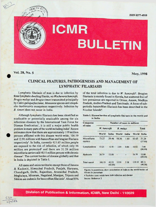
in India is depicted in Table I.
Table I. Disease burden of lymphatic filariasis in the world and
in India.
All states and union territories except those of Jammu
,& Kashmir, Himachal Pradesh, Punjab, Haryana,
Chandigarh, Delhi, Rajasthan, Arunachal Pradesh,
Meghalaya, Mizoram, Nagaland, Manipur, Tripura and
Sikkim are endemic for bancroftian filariasis2. About 95%
Categories
of cases
Number of cases in millions
W. bancrofti
B. malayi
Total
World
India
World
India
World
India
Microfilaria
carriers
73.27-
29.46
10.36
1.80
83.63
31.26
(37.00)
Lymphoedema
13.21
6.58 -
2.81
0.86
16.02
7.44
(46.40)
Hydrocele
26.79
12.88
-
-
26.79
12.88
(48.00)
Total cases#
106.19
45.53
12.91
(42.88)
2.53
119.10
48.11
(40.30)
Figures in parentheses show contribution of India to the world burden of
disease in percentage.
# Excludes cases which have both infection and disease
Source: Reference 3
Division of Publication & Information, ICMR, New Delhi - 110029
This disease due to its prolonged course and non
fatal nature has attracted little attention of the health plan
ners, thus a low priority has been accorded by the States
to the National Filaria Control Programme5. The disease,
however, has serious socio-economic impact on the so
ciety by the significantly lowered productivity and eco
nomic loss resulting from its chronicity6*11. In addition,
there are stigmatizing effects due to the grotesque dis
figurements as sequelae of the disease. The number of
individuals world-wide with overt physical disabilities
resulting from the infection is approximately 43 million,
with bancroftian filariasis accounting for about 40 mil
lion of these cases12. Lymphatic filariasis is the second
leading cause of permanent physical disability in humans
next to mood disorders13.
The public health impact of different diseases is com
pared by using Disability Adjusted Life Years (DALYs)
as a standardized metric by the World Bank Develop
ment Report (WDR)14. By using current estimates of the
disease incidence with focus on chronic manifestations
only, the global burden of lymphatic filariasis by this
measure has been estimated by WDR to be 850,000 DALYs
lost representing a mere 0.23% of the total global bur
den of parasitic and infectious disease. This is seen as a
gross underestimate of the disease incidence which ig
nores the influence of acute disease on DALYs4-6. It has
been estimated that on an average 23.4 and 26.5 mandays are lost per individual per year due to bancroftian
and brugian filariasis respectively and an overall 162.12
million man-days are lost per year in India due to acute
filarial attacks15. If the cost of treatment including hos
pitalization and surgical procedures were also to be in
cluded, the actual loss would have been manifold. Acute
disease is likely to be many times more prevalent than
the chronic16. In a recent study in an endemic rural area
of Tamil Nadu in south India, it was shown that the mean
cost incurred on treatment of each episode of the acute
disease ie. adenolymphangitis (ADL) amounted to Rs.
2.35. It was further estimated, that the cost of treatment
of the acute disease (per attack of ADL) was equal to the
wages for. 2 days for men and for 4 days for women17.
Presently, filariasis control does not only imply inter
ruption of disease transmission, but also morbidity con
trol by appropriate medical or surgical procedures. In
addition, there has of late been a sea change in our un
derstanding of the pathogenesis and progression of the
disease process by hitherto unavailable diagnostic tools
like ultrasound18 and lymphangioscintigraphy19*21. The defi
nition of the infected person itself has taken a U-turn,
since the individual without symptoms or microfilaria
42
in a filaria endemic zone can no longer be corisidered
normal following direct visualization of the parasite by
the use of these new diagnostic technologies and by
the detection of renal and lymphatic abnormalities in
apparently normal individuals. The principles of mass
as well as individual chemotherapy have also witnessed
a dramatic change. The current review encompasses these
recent advances in the understanding of lymphatic
filariasis.
Clinical Features
The clinical manifestations of lymphatic filariasis tend
to vary in different geographical locations2243. The dis
ease spectrum of bancroftian and brugian filariasis var
ies widely from an asymptomatic state to a severely
debilitating chronic condition. Depending upon the in
fection status of the individuals in endemic areas and <
the resulting clinical consequences thereof, five main
categories of the filarial clinical spectrum have been
recognized4. These are: (i) asymptomatic amicrofilaraemia;
(ii) asymptomatic microfilaraemia; (iii) acute manifes
tations; (iv) chronic manifestations; and (v) allergic mani
festations.
Besides, there are reports of manifestations like glo
merulonephritis and monoarthritis rn patients with
bancroftian filariasis24*27.
Asymptomatic Amicrofilaraemia
A proportion of individuals in endemic areas neither
show any microfilaria (mf) in the blood nor exhibit any
symptoms. This may comprise persons who have not been
exposed enough to become infected or persons though
sufficiently exposed, do not show any detectable infec
tion by current diagnostic techniques4. It may be that they
have successfully cleared the infection (immune indi
viduals) or are harbouring developing worms of one sex
or unfertilized female adult worms. This group may have
subclinical infection as evidenced by detectable
antigenaemia28*34.
Asymptomatic Microfilaraemia
Some individuals who develop .microfilaraemia do
not manifest any clinical disease. Among these, some
may remain asymptomatic for years even for life, while
others manifest clinical disease with or without losing
the microfilaraemic status4. Besides, this group of indi
viduals shows lowered parasite specific IgE response but
generate large amounts of specific antibody of the IgG4
class which is considered as a blocking antibody33*3*.
Acute Manifestations
9
The acute manifestations are characterized-by epi
sodic attacks of ADL with constitutional symptoms like
fever, chills, malaise, headache, nausea and vomiting43637.
The ADL attacks usually involve the limbs and the groins
and also the male genitals (acute funiculitis or epididymitis
or orchitis, most commonly involving all three simulta
neously), the female breast and rarely other sites. Acute
attacks never occur at more than one site at the same
time36. In case of the limb, groin or breast, there are local
signs of lymphangitis and lymphadenitis (redness, swelling,
raised local temperature, pain and tenderness at the lo
cal site). These attacks have been described as
adenolymphangitis, since the patients usually complain
of lymph node involvement followed by retrograde lym
phangitis36,38. Sometimes infection of deep lymphatics
may mimic an acute abdominal emergency as if to
require surgical intervention immediately39,40.
The ADL attacks commonly occur in patients with
established chronic filarial disease, more frequently with
lymphoedema than with hydrocele34. The attacks can also
occur in asymptomatic persons, many of whom present
with lymphoedema with the first attack itself. Most of
ten, repeated attacks of ADL preceqe the development
of chronic lymphatic pathology of filariasis and these
often continue for many years34,41. The ADL attacks usu
ally last for about 3 to 5 days, but rarely may be pro
longed upto a fortnight10,1637,41. The frequency of these
attacks is usually 1 to 3 in a year. However, it can also
be more than twice a month in some patients16. Each attack
of fever and lymphadenitis subsides spontaneously with
a crisis. Lymphoedema is frequently present during these
episodes, but usually subsides after the acute stage. With
time, resolution of the lymphoedema after each attack
becomes less complete and the characteristic chronic
^changes develop42.
Chronic Manifestations
The common chronic manifestations of lymphatic
filariasis affect the male genitals and limbs of both sexes.
Most of these patients are amicrofilaraemic39,43-46. The
incidence and severity of these chronic clinical
manifestations tend to increase with age4,47,48. These
manifestations are the result ofstructural damage following
lymphatic obstruction and fibrosis. In brugian filariasis,
genitals are rarely involved.
Hydrocele
Hydrocele is the commonest manifestation of chronic
bancroftian filariasis. It has only been very rarely recorded
in brugian filariasis. It presents as a swelling of the peri
toneal lining that surrounds each of the testicles. A clear
str^w coloured fluid accumulates in the closed sac (hy
drocele) as a result of the blockage in the lymphatics
draining the retro-peritoneal and sub-diaphragmatic ar
eas. Rarely the fluid has a milky appearance caused by
the presence of chyle, a condition known as chylocele.
Sometimes mf can be detected in the hydrocele fluid4,49.
Besides hydrocele, chronic epididymitis, funiculitis, lymph
scrotum (vide infra) are other genital manifestations of
chronic bancroftian filariasis in males4. Though in an
endemic region hydrocele is assumed to be of filarial
origin and conventionally used as an absolute indicator
of filarial disease in clinical surveys,.even in known
endemic areas of India, eg., Pondicherry and Orissa, only
69 and 43% of cases of hydrocele respectively could be
demonstrated to be of definite filarial etiology?930. Simi
lar findings have been reported from Puerto Rico51.
Lymphoedema
For both sexes, and independent of age, it has been
observed that approximately 11% of the population at
risk eventually develop lymphoedema. Lymphoedema
commonly affects the lower limbs, rarely upper limbs
and genitals in both sexes and female breast. In both forms
of lymphatic filariasis, the legs are most commonly in
volved52*54. Lymphoedema can be graded as follows55:
Grade I
: Oedema spontaneously reversible on elevation.
Grade II : Oedema not spontaneously reversible on
elevation, skin normal.
Grade III: Oedema not spontaneously reversible on
elevation, skin thickened.
Grade IV: Oedema not spontaneously reversible on
elevation, skin thickened with warty/papillomat'ous growth.
Elephantiasis supervenes following persistent fibrotic
changes in the skin and subcutaneous tissue. Elephan
tiasis can also affect the genitals as stated above (lymph
scrotum or penis in males and vulva in females) and the
female breast4,39.
Chyluria
Chyluria is the excretion of milky urine due to ad
mixture of chyle with urine. A minority of the affected
subjects complain of gross haematuria. The condition is
due to the blockage of the retro-peritoneal lymph nodes
below the cistema chyli, with consequent reflux and flow
43
of the intestinal lymph directly into the renal lymphatics
which may rupture and permit flow of chyle into the urinary
tract. Microfilaraemia may or may hot be present in these
patients. The urinary sediment may contain microfilaria
and red cells but seldom leucocytes. Chyluria is often
symptomless, but some patients complain of fatigue and
weight loss due to loss of fat and protein4-39,56.
Allergic Manifestations
Tropical pulmonary eosinophilia (TPE) is a form of
occult filariasis developing in a small proportion of in
dividuals in areas endemic for filariasis. It is considered
as an allergic manifestation of the disease with extremely
high titres of anti-filarial antibodies4. A majority of these
antibodies is of IgE class and is directed against microfi
laria57. Hypereosinophilia is the most common feature
with absolute eosinophil counts ranging from 3,000-50,000
cells/mm3 of blood. Here the classical clinical manifes
tations are not present, with absence of circulating mf in
the blood, but probably present in the tissues particu
larly the lung. The commonest presentation is nocturnal
paroxysmal cough with radiological evidence of diffuse
miliary lesions or increased broncho-vascular markings
and elevated erythrocyte sedimentation rate4. There is
impaired lung function in most cases with a reduction in
the vital capacity, total lung capacity and residual vol
ume58. Males are affected twice as often as females and
the disease is rarely reported in children4. The therapeu
tic response to diethylcarbamazine (DEC) is generally
good. If untreated (even in some treated cases) TPE may
progress to chronic pulmonary fibrosis58.
Differences between Bancroftian and Brugian Filariasis
In India, bancroftian and brugian filariasis are
characterized by a wide range of clinical manifestations
presented by only a small proportion of infected indi
viduals.
Although, it has been reported that in bancroftian
filariasis genital involvement is more in the north Indian
states when compared to states in south India22, subse
quent studies have shown that the disease distribution is
more or less uniform44,59. The reported discrepancy was
attributable to the difference in methods of clinical ex
amination and improper survey design48. In periodic
brugian filariasis in Kerala, the clinical spectrum of the
disease though found to be generally similar to that re
ported elsewhere in the world,6,36,60 the progress of oedema
to elephantiasis was observed to be a slow process ex
tending even upto a period of 20 years probably related
44
to the frequency of ADL attacks53, and abscess forma
tion in the inguinal lymph nodes and along the main lym
phatic channel was found to be rare53 though reported to
be common in similar infection in Indonesia52 and Sri
Lanka6. This is in contrast to bancroftian infection in south
India where elephantoid changes and dermatosclerosis
appear more rapidly53. In brugian filariasis, involvement
of the male genitals was found to be much less common
as compared to bancroftian filariasis in Kerala53 as found
elsewhere6,36. A comparison between the clinical aspects
of the two forms of lymphatic filariasis is shown in Table
II.
Table II. Comparison of clinical aspects of bancroftian and brugian
filariasis.
Bancroftian filariasis
Brugian filariasis
Progression
of the disease33 •
Fast
Relatively slow
Relation with
gender3’-4*-53
Occurrence in males
more than in females
No difference
between genders
4.9±1.70
5.5±1.01
10.4±3.20
0.9±0.22
2.5±0.29
-5.1*1.08
Grade I
Grade II
Grade III
0.1±0.02
5.8±0.56
8.6±0.92
0.1 ±0.01
13.6*1.17
28.7±2.66
Involvement of
male genitals39-33
Common
Rare
Extent of elephantiasis39
Above kneee
Parameter
Frequency of
acute attacks per
year in patients
with lymphoedema34
Grade I
Grade II
Grade III
Mean duration (yr)
of lymphoedema34
Below kneee
___________________ A
Superscript numbers refer to sl.nos. in the reference list
Filarial arthritis which may manifest in endemic
regions is monoarthritic usually affecting the knee joint.
Other conditions suggested to be associated with
lymphatic filariasis include endomyocardial fibrosis,
glomerulonephritis, thrombophlebitis, tenosynovitis, nerve
palsies, dermatoses, etc. Lymphoedema has also been
considered to predispose to lymphosarcoma61.
Pathogenesis
Observations on studies undertaken in Pondicherry
and Calcutta suggest that filariasis endemic populations
consist of individuals who remain amicrofilaraemic and
asymptomatic and those who progress through the se
quence : uninfected, microfllaraemic and amicrofilaraemic
to develop irreversible lymphatic .pathology62.
Conventionally, it had been the view that the patho
genesis of the lymphatic abnormalities in this disease is
essentially mediated by the immune system of the host33
but the advent of recent techniques such as ultrasound"
and lymphangioscintigraphy1’’21 has shown that factors
other than immunological, such as the parasite itself may
play a role in the pathogenesis. The presence of the adult
worms in the lymphatics causes early lymphatic dysfunc
tion and dilatation etc due to factors released by the parasite
as described in other systems63 or elaborated directly by
the host. The primary role of the immune system in this
early lympathic dysfunction appears to be the produc
tion of down-regulatory molecules which contain or inhibit
inflammatory responses64; this hypothesis being supported
Q by recent findings that cytokine responsiveness in
microfilaraemics is of the Th-2 type ie. heightened pro
duction of interleukins -4, -5 and -10 which are contrainflammatory 65-“. Th-2 type down regulating suppres
sive cytokines in asymptomatic microfilaraemics facili
tate persistence, of the parasite in the host while induc
ing little or no inflammatory clinical disease. IL-4 stimu
lates B cells proliferation resulting in IgG4and IgE pro
duction where IgG4 acts as blocking antibody with con
sequent reduction of the inflammatory activity of IgE2’.
This contrasts with the relatively pro-inflammatory
Th-1 response seen in amicrofilaraemic patients with
chronic lymphatic pathology following breakdown of this
tolerance. This is associated with a high level of IgE,
IgG] and IgG, and low level of IgG4. Elephantiasis may
result by IgE mediated mechanisms or a passive reac
tion to naturally dying adult worm evoking IgG, and IgG2
antibody response mediating inflammatory reactions. This
immunological hyper-responsiveness may remove the
parasite from circulation as in developing lymphoedema67.
Though microfilaraemics remain asymptomatic for
long periods, they cannot be considered to be truly free
from any abnormalities. The asymptomatic state earlier
thought to be benign is no longer so; it has been demon
strated that most of the so called asymptomatic
microfilaraemics have lymphatic abnormalities as detected
by lymphangioscintigraphy and also renal abnormalities
as evidenced by microscopic haematuria and/or proteinuria.
Renal abnormalities are generally reversible after treat
ment27. Prenatal tolerance to filarial antigens has been
shown to determine immune responsiveness to the
parasite and thereby the development of lymphatic pa
thology ie. children bom of microfllaraemic mothers are
much less capable of responding to filarial antigens than
those bom of non-infected mothers6*-6’. However, reefent
studies on bancroftian microfilaraemia in children in re
lation to parental infection have shown that there is hardly
any role of intrauterine sensitization, since the
microfllaraemic status of the father has also been seen
to influence microfilaraemia in bancroftian infection of
the children70. This only highlights the influence of house
hold exposure on all family members.
The findings in animal models parallel those in the
affected humans, ie. lymphatic proliferation, dilatation
and oedema formation due to the presence of living adults
and obstructive, obliterative reactions in the lymphatics
around dead parasites. The earliest changes occur four
weeks after infection and involve the regional lymph nodes.
Within four days of the infecting larvae reaching the
lymphatics, there is a marked cell mediated response in
the regional lymph nodes followed by an antibody me
diated response in the afferent lymphatics. The local lymph
node is enlarged followed by an increase in diameter and
formation of tortuosities of the lymphatics through which
the infective larvae migrate. These changes may revert
if the worms are killed by microfilaricidal drugs71. Fol
lowing appearance of antimicrofilarial antibodies, resis
tance to reinfection develops causing obstruction to the
lymph flow resulting in lymphoedema. Initially there is
an acute inflammatory response characterized by infil
tration of polymorphs, histiocytes, eosinophils and lym
phocytes around the lymphatics. This is followed by
formation of epitheloid granuloma and foreign body giant
cells after the death of the adult worms, which may cal
cify, become lysed and surrounded by fibrosis. Lymphatic
abscesses may form at the site of dead and degenerating
worms. Finally, the lymphatics are obliterated by fibro
sis with the disappearance of microfilariae from the blood71.
Lymph stasis results in increased susceptibility to sec
ondary bacterial infection particularly with group A strep
tococci resulting in episodic attacks of ADL. Each at
tack of ADL results in the progression of chronicity of
disease and increased lymph stasis, thereby, again in
creasing susceptibility to ADL attacks16. This culminates
in fibrosis with the formation of solid oedema and prob
able decline in ADL attacks.
The pathogenesis of late stage abnormalities (lym
phoedema and elephantiasis) includes an inflammatory
component comprising significant cellular infiltration in
the epidermis and hypodermis as revealed by examina
tion of biopsy specimens of skin from patients of filarial
lymphoedema. In comparison, similar examinations in
individuals with non-filarial lymphoedema showed far
45
fewer changes72. Alternatively, in a compromised state
of lymphatic function, superinfection of tissue with micro
organisms may be largely responsible for local inflam
mation67 and eventual elephantiasis as the clinical out
come of this disease73.
antihistaminics may be used if indicated. Symptomatic
treatment for nausea and vomiting if necessary can be
given. Immobilization and elevation of the affected limb
should be carried out to ameliorate suffering during the
acute attack. In case ofabscess formation, surgical drainage
is to be done with necessary follow up.
Management
Standard regimen of DEC is to be administered after
the acute attack of ADL has subsided to avoid severe
allergic reactions during the acute attack.
The management of lymphatic filariasis varies ac
cording to the stage of infection, disease manifestations
and its complications. The overall management comprises
mainly medical measures and surgical intervention is only
called for if indicated. Medical measures include
physiotherapy and chemotherapy for the acute and chronic
forms of the disease. The principles of management
accordingly are enunciated below:
Asymptomatic Infected
Chemotherapy is the mainstay of treatment of mf car-t
riers and antigen positive individuals. The following drug
or combination of drugs are in use currently : diethylcarbamazine citrate (DEC) is the drug of choice. It-is
used singly or in combination with other anthelmintics
eg. ivermectin or albendazole. One ofthe following sched
ules is advocated, (i) DEC: 6 mg/kg body weight (b w)
in three divided doses per day after meals for 12 days
[W.bancrofti] or for 6 days [B. malayi] - standard regi
men12, or (ii) DEC: 6 mg/kg b w in single dose at monthly
intervals for 12 months, or (iii) Ivermectin: 400 gg/kg
b w repeated at three monthly intervals for 12 months
(not marketed currently in India for human use), or
(iv) DEC and ivermectin: a single dose of DEC 6 mg/kg
b w and ivermectin 200 pg/kg b w repeated after 6 months,
or (v) DEC and albendazole: DEC 6 mg/kg b w and
albendazole 600 mg for adults to be repeated after one
year if required74.
Acute Manifestations
Antibiotic therapy has, recently gained importance
following recent studies that have demonstrated the role
of bacteria especially beta haemolytic streptococci in the
causation of ADL episodes67. Penicillin, the drug of choice,
is given as Benzathine penicillin G 1,200,000 units by
injection every 4 weeks or penicillin G potassium salt
250,000 units or 250 mg penicillin V orally 3 to 4 times
a day till the symptoms subside75. Synthetic penicillin
analogues like amoxycillin can also be given. If allergic
to penicillin, erythromycin or other macrolides may be
used. Antipyretics eg. paracetamol; non-steroidal anti
inflammatory drugs (NSAID) eg. diclofenac sodium and
46
Chronic Manifestations
Hydrocele
Most hydroceles of small size may not require any
treatment. For moderate to large hydroceles, the treat
ment of choice is surgical. Monthly courses of DEC
(10 mg/kg b w per day for three weeks) for a period of
6 months have been reported to be beneficial76. How
ever, further evaluation in this regard is required.
Lymphoedema
Management of lymphoedema calls for both prophy
lactic and therapeutic measures. Therapeutic measures
comprise the following:
(i) Medical
(a) Physiotherapy: It is useful in the early stages of
lymphoedema and includes the use. of pressure
bandage, manual massage, thermotherapy, pneumatic
compression and interferential current therapy.
Pressure bandage: Crepe bandages and elasto-crepe
stockings are commonly used and the latter are pre
ferred for simplicity, but they have to be tailor made
for individual patients. Care has to be taken to edu- WP
cate the patients in the proper use of these, since these
are often used inadequately and improperly.
Manual massage: The principle is to mobilize the
oedema fluid away from the affected part milking it
upwards along the cutaneous and subcutaneous net
works to the abdomen. Though the experience in
filarial lymphoedema is limited, it has the potential
for domiciliary management since the patients and
relatives could be educated and trained in this therapy
for daily application. It is a technique where gentle
pressure is applied on the skin to reduce the pain and
oedema of the affected region by promoting lymph
circulation from the periphery to the centre. The mas
sage needs to be done daily and requires a lot of
patience and motivation on the part of the patient as
well as total involvement of the therapist. The mas
sage needs to be done daily and evaluated regularly
at monthly intervals. The details of the procedure
are given elsewhere74.
Thermotherapy: It is useful both for the reduction of
oedema and the associated pain. Wet heat therapy
by immersing the affected limb in a bucket of warm
water for about 10 to 15 min daily in the evenings
when the size of the swelling is increased due to the
day’s activities, is recommended to facilitate circu
lation. After this, the patient could massage the limb
and lie flat with the foot being kept elevated. In ad
dition, hot ovens (modified Chinese therapy) have
also been found to be useful.
Pneumatic compression: Single and multi-cell jack
ets are used for pneumatic compression of affected
parts either by intermittent low pressure or continu
ous high pressure generated by electrically operated
machines. Reduction occurs immediately after therapy,
but the results are not sustained in course of time
particularly in ambulatory patients. Hence, elastic ban
daging as an adjunct after compression is essential.
Interferential current therapy: Low frequency inter
ferential current stimulation is given to the affected
part using two electrodes placed diametrically op
posite on the skin, with one having a fixed stimula
tion (4000 Hz) and the other variable (3850 to 4000
Hz). This creates an interferential beat frequency
equivalent to the difference between the two through
the tissue. This therapy though tried on a limited scale
has been found to be useful in the reduction of pain
and oedema particularly in patients with grades II
and III lymphoedema (VCRC data).
(b) Chemotherapy; Standard regimen of DEC as for mf
carriers can be given every month for .12 months. Apart
from this coumarin 200 mg twice a day has been re
ported to be helpful in the reduction of filarial oedema
in the long run. However, the role of coumarin needs
further evaluation.
(ii) Surgical interventions
In lymphoedema, surgery is resorted to only in cases
who do not respond to aggressive medical management
or have impaired functions particularly in cases with
physical and occupational disabilities or for social reasons. The surgical procedures include lymphatic drain
age and/or excisional surgery.
(a) Drainage procedures: These include lymphangioplasty,
omentoplasty, flaps, lymph-vein anastomosis, lymph
lymph anastomosis, lympho-nodo-venous (LNV)
shunt, etc. LNV shunt shows immediate good response
epecially in patients with moderate to high oedema
ie. grade III with oedema volume more than 2 litres
and duration of about 5 years. There is a rapid re
duction in oedema volume.(about 40%). The gains
are not sustained and in most ofthe patients the oedema
volume gets gradually increased. Post-surgery ADL
attacks are the most important cause for the failure
of the viability of the shunt. However, LNV shunt
may have a role as a pre-excisional surgical proce
dure in filarial lymphoedema
(b) Excisional procedures: These procedures are resorted
to only in patients with gross physical deformities
or occupational disabilities. Reliable objective as
sessment of excisional procedures are not available
for long-term results.
Medical management of lymphoedema is of primary
importance since it can be undertaken at domiciliary, PHC
or referral levels. Surgery is advocated in selected cases
as a last resort since it can be accomplished in referral
centres only. Though the long-term value of any proce
dure is still not certain, the cases need to be followed up
by objective limb circumference measurement at fixed
points.
Allergic Manifestations
Tropica! pulmonary eosinophilia, a form of occult
filariasis considered as the most important allergic
manifestation of this disease, responds fairly well to
standard regimen of DEC. If neglected, it may lead to an
irreversible condition of chronic pulmonary fibfosis.
However, relapses may occur in 20%’ of patients who
may require higher doses of the drug ie. 6-12 mg/kg b w
daily for 21-30 days. Beneficial effects of DEC have
also been observed in other forms of occult filariasis.
Management ofAdverse Reactions to Anti filarial Drugs
The adverse reactions to ^ntifilarial drugs may re
sult in two ways. The first is a pharmacological dose
dependent response to the chemical properties of the drug
itself. This may occur in infected as well as in uninfected
recipients of the drug with equal frequency. The second
is due to the response of the infected host to the destruction and death of the parasite and is independent of the
dose but is related to the pre-therapy parasite load77'79.
Usually, there is hardly any adverse effect due to phar
macological toxicity of the standard dose of DEC in the
uninfected person. At higher dosage only, there may be
.47
anorexia, nausea, abdominal pain, weakness, dizziness
or lethargy which may begin 1 72 hours after ingestion of
the drug on an empty stomach and persist for a few hours.
There is no accumulation of the drug following prolonged
administration and chronic toxicity does not occur. The
second type of reactions is attributed to the filaricidal
action of DEC. These reactions are the result of immu
nological response to the disintegrating microfilariae and
dead adult parasites. They are usually less severe in
bancroftian than in brugian filariasis. These reactions may
be systemic or local, both with and without fever.
Systemic reactions include malaise, headache, body
ache,joint pain, dizziness, anorexia, vomiting, transient
haematuria, allergic reactions and rarely bronchial asthma.
They usually occur a few hours after the first dose of
DEC and last for about 3 days. Fever may be present.
The systemic reactions cease spontaneously and symp
tomatic treatment with antipyretics and analgesics is givert.
Local reactions usually comprise lymphadenitis, ab
scess, ulceration, transient lymphoedema and hydrocele
occurring in decreasing frequency. In bancroftian filari
asis, there may be funiculitis and epididymitis. Though
they appear later in the course of the disease and last
longer, they also disappear spontaneously and interrup
tion of treatment may not be necessary. In cases where
lymphoedema and hydrocele have persisted for several
months after DEC therapy, elephantiasis has not been
observed. Local reactions tend to occur more often in
patients with history of ADL being probably related to
the presence of adult or immature worms or fourth stage
larvae in the tissues.
Prophylaxis of Filariasis
For prevention and management of the acute disease,
foot hygiene is of prime importance74. This consists of
the following : (i) cleaning feet with soap containing 2%
glycerine, (ii) regular application of Whitfield ointment
on the webs of toes to prevent fungi infection and to
smoothen the skin, (iii) regular clipping of toe and fin
ger nails. Clipping of finger nails is important to prevent
injuring the skin of the affected limb by scratching on
itching, and (iv) prompt and appropriate attention to all
injuries in the body specially the feet.
Other measures of prevention include (i) treatment
of any other focus of infection eg. caries tooth, CSOM
etc.; and (ii) for patients who get frequent attacks of ADL,
long acting penicillin (benzathine penicillin 12 lakhs units)
intra-muscular once every 3 weeks after carrying out man
datory sensitivity skin test or oral penicillin V 100 00
48
HJ, 65 mg tablet can be given daily (VCRC unpunished
data).
As stated above, in endemic areas, individuals termed
endemic normals may not be so, since the absence of
symptoms or microfilaraemia does not necessarily mean
that they are uninfected. In a proportion of this popula
tion, infection may be evidenced only by the demonstra
tion of antigenaemia, whereas in the rest, the presence
of infection or otherwise cannot be ascertained by the
currently available means. This justifies the institution
of mass chemotherapy for the infected and individual
prophylaxis for the supposedly uninfected by antifilarial
drugs.
In endemic areas, preventive measures against mos
quito bites eg. bednet and mosquito proofing of the house
should be taken. In addition, DEC 300 mg as a single
dose after dinner per day for 2 days every month acts as
prophylaxis against filariasis for people from non-endemic
regions visiting endemic regions immediately on arrival.
In an area with cofirmed active transmission of filariasis, DEC at a dosage of 500 mg on each of two con
secutive days each month for 18 months has been shown
to be prophylactic throughout and also for further 12
months after the cessation of DEC administration80.
Conclusions
Although lymphatic filariasis continues to be a ma
jor public health problem in many parts of the world
particularly India, it has of late been identified as a po
tentially eradicable disease. The clinical features of lym
phatic filariasis present a highly variable spectrum and
recent advances in diagnostic techniques have led to
questioning of the earlier concept of endemic normals,
since lymphatic and renal abnormalities have been directly visualized in otherwise apparently normal indi
viduals in endemic regions. In such cases as also in ar
thritis associated with this disease, the exact etiopathogenesis is not fully understood. Control of acute attacks
of ADL by antibiotics and simple measures like foot
hygiene etc. have been shown to prevent the progression
of the disease to its chronic, from with the resultant dis
ability. Though DEC continues to be the mainstay of
individual as well as mass chemotherapy, newer
antifilarials like ivermectin and anthelmintics like
albendazole are being added to the arsenal of antifilarial
treatment. The earlier sense of hopelessness in morbid
ity management has also given way to a new sense of
optimism and hope both to the clinician and the patient
who can be given simple training of self support.
e
Lymphatic abnormalities in human filariasis as depicted by
lymphangioscintigraphy. Arch Int Med 153: 753, 1993.
References
1.
Centre for Disease Control. Recommendations of the Interna
tional Task Force for Disease Eradication. Morb Mortal Wkly
Rep 42 (RR-16): 1, 1993.
National Filaria Control Programme Operational Manual. Di
rectorate, National Malaria Eradication Programme, Delhi, 1995.
20.
Freedman, D.O., Filho, P.J., Besh, S., Silva, M., Braga, C. and
Maciel, A. Lymphangioscintigraphic analysis of lymphatic ab
normalities in symptomatic and asymptomatic human filariasis.
J Infect Dis 170: 927, 1994.
21.
Dreyer, G., Figueredo-Silva, J., Andrade, L.D., Marchetti, F. and
Coutinho, A. Diagnosis, treatment and control of filariasis. Parasite
1 (Suppl 1): 22, 1994.
22.
Hawking, F. The distribution of human filariasis throughout the
world. Part II. Asia. Trop Dis Bull 73: 967, 1977.
23.
Rao, C.K. Current knowledge on selected aspects of the epide
miology of bancroftian filariasis. J Commun Dis 9: 185, 1977.
24.
Chaturvedi, P, Gaw'di, A. and Dey, S. Occult filarial infections.
Natl Med J India 3: 7, 1990.
25.
Jamal, S: and Pani, S.P. The clijiical perspectives and research
needs in lymphatic filariasis. (Proceedings of a Seminar on Fu
ture Research Needs in Lymphatic Filariasis). VCRC Pondichery,
Misc Publ No. 16: 29, 1990.
26.
Rath, R.N., Mishra, N., Sahu, R.N., Mohanty, G. and Das, B.K.
Renal involvement in bancroftian filariasis. Natl Med J India 4:
65, 1991.
27.
Dreyer, G., Ottesen, .A., Galdino, E., Andrade, L., Rocha, A.,
Medeiros, Z., Moura, L, Casimiro, L, Beliz, F. and Coutinho, A.
Renal abnormalities in microfilaraemic patients with bancroftian
filariasis. Am J Trop Med Hyg 46: 754, 1992.
3.
Michael, E., Bundy, D.A.P. and Grenfell, B.T. Reassessing the
global prevalence and distribution of lymphatic filariasis. Para
sitology 112: 409, 1996.
4.
World Health Organization. Lymphatic filariasis: The disease
and its control. WHO Tech Rep Ser 821: 1992.
5.
Rao, C.K. and Sharma, S.P. Control of filariasis in India. JCommun
Dis 18: 276, 1986.
6.
Raghavan, N.G.S. Clinical manifestations and associated epide
miological factors of filariasis. J Commun Dis 1: 75, 1969.
7.
Ramaiah, K.D., Vijayakumar, K.N., Ramu, K., Pani, S.P. and
Das, P.K. Functional impairment caused by lymphatic filariasis
in rural areas of south India. Trop Med Int Health 2: 832, 1997.
8.
Ramu, K., Ramaiah, K.D., Guyatt, H. and Evans, D. Impact of
lymphatic filariasis on the productivity of male weavers in a south
Indian village. Trans R Soc Trop Med Hyg 90: 669, 1996.
9.
Gyapong, J.O., Gyapong, M., Evans, D.B., Aikins, M.K. and
Adjei, S. The economic burden of lymphatic filariasis in north
ern Ghana. Ann Trop Med Parasitol 90: 39, 1996.
10.
Belizario, V., Lariosa, T., Pesigan, A., Leonardia, W. and Llanto.
The clinical epidemiology of bancroftian filariasis in an endemic
village in Sarsogon, Philippines. Acta Medica Phililpina 31: 61,
1995.
28.
Bundy, D.A.P., Grenfell, B.T. and Rajagopalan, P.K.
Immunoepidemiology of lymphatic filariasis: The relationship
between infection and disease. Immunol Today 12(3): A71,1991.
11.
Evans, D.B. Gelband, H. and Vlassoff, C. Social and economic
factors and the control of lymphatic filariasis. A>review. Acta
Tropica 53: I, 1993.
29.
Maizels, R.M. and Lawrence, R.A. Immunological tolerance: The
key feature in human filariasis. Parasitol Today 7: 21, 1991.
12.
World* Health Organization. Lymphatic filariasis infection and
disease: Control strategies. Rep TDR/CTD/FIUPenang/94.1/1994.
30.
Day, K.P. The endemic normal in lymphatic filariasis: A static
concept. Parasitol Today 7: 341, 1991.
13.
The World Health Report: Bridging the Gap. Report of the Di
rector-General, World Health Organization, Geneva, p 3, 1995.
31.
14.
World Bank. World Development Report: Investing in Health
(WorldDevelopment Indicators). Oxford University Press, New
York, p 25, 1993.
Grenfell, B.T., Michael, E. and Denham, D.A. A model for the
dynamics of human lymphatic filariasis. Parasitol Today 7:318,
1991.
32.
Sabesan, S., Krishnamoorthy, K., Pani, S.P. and Panicker, K.N.
Man-days lost due to repeated attacks of the acute disease of
lymphatic filariasis. Trends Life Sci 7: 5, 1992.
Grenfell, B.T. and Michael, E. Infection and disease in lymphatic
filariasis - an epidemiological approach. Parasitology 104 (Suppl):
S81, 1992.
33.
Ottesen, E.A.* Infection and disease in lymphatic filariasis.
An immunological perspective. Parasitology 104 (Suppl): S71,
1992.
34.
Pani, S.P. and Dhanda, V. Natural history and dynamics of pro
gression of filariasis. In: Proceedings ofthe CSIR Golden Jubi
lee Symposium on Tropical Diseases, Molecular Biology and
Control Strategies. Eds. S. Kumar., A.K. Sen., G.P. Dutta, and
R.N. Sharma, Publication and Information Directorate, New Delhi,
p.l, 1994.
35.
Amaral, F., Dreyer, G., Figueredo-Silva, J., Noroes, J., Cavalcanti,
A., Samico, S.C., Santos, A. and Coutinho, A. Live adult worm
detected by ultrasonography in human bancroftian filariasis. Am
J Trop Med Hyg 50: 753, 1994.
Ottesen, E.A. Filariasis now. Am J Trop MedHyg 41 (Suppl 3):
9, 1989.
36.
Turner, L.H. Studies on filariasis in Malaya: The clinical fea
tures of filariasis due to Wuchereria malayi. Trans R Soc Trop
Med Hyg 53: 154, 1959.
Witte, M.H., Jamal, S., Williams, W.H., Wittee, C.L.,
Kumaraswami, V., McNeil, G.C., Case, T.C. and Panicker, T.M.R.
37.
Weller, P.F. Paroxysmal inflammatory filariasis: filarial fevers.
Arch Int Med 143: 1523, 1983.
15.
16.
17.
18.
19.
Pani, S.P., Yuvaraj, J., Vanamail, P., Dhanda, V., Michael, E.,
Grenfell, B.T. and* Bundy, D.A.P. Episodic adenolymphangitis
and lymphoedema in patients with bancroftian filariasis. Trans
R Soc Trop Med Hyg 89: 72, 1995.
Ramaiah, K.D., Ramu, K., Guyat, H., Vijaykumar, K.N. and Pani,
S.P. Direct and indirect costs of the acute form of lymphatic
filariasis to households in rural areas of Tamil Nadu, south
India. Trop Med Int Health 3: 108, 1998.
49
38.
Kumaraswami, V. Clinical aspects of filariasis. In: Proceedings
of the First International Conference on the Control of Lym
phatic Filariasis. Ann Austral College Trop Med Inc.' 2: 39,
1997.
39.
Partono, F. The spectrum of disease in lymphatic filariasis. In:
Filariasis. CIBA Foundation Symposium, No. 127. John Wiley
& Sons, Chichester, U.K. p 15, 1987.
40.
Wartmap, W.B. Filariasis in American armed forces in World
War II. Medicine 26: 333, 1947.
41.
Ramaiah, K.D., Ramu, K., Vijaykumar, K.N. and Guyatt, H.
Epidemiology of acute filarial episodes caused by Wuchereria
bancrofti infection in two rural villages in Tamil Nadu, south
India. Trans R Soc Trop Med Hyg 90: 639, 1996.
42.
Dissanaike, A.S. Filarial infections. In: Recent Advances in Tropi
cal Medicine. Ed. H.M. Gillies. Churchill Livingstone, Edinburg
p.115, 1984.
55.
World Health Organization. Informal consultation on evaluation
of morbidity in lymphatic filariasis. WHO/TDR/FIL Mimeo
graphed Document: 92.3,-1992.
56.
Das, S. and Dandapat, M.C. Management of acute manifesta
tions of filariasis. J Indian Med Assoc 73: 215, 1979.
57.
Ottesen, E.A. Immunological aspects of lymphatic filariasis and
onchocerciasis in man. Trans R Soc Trop MedHyg 78: 9, 1984.
58.
Vijayan,'V.K., Kuppu Rao, K.V., Sankaran, K., Venkatesan, P.
and Prabhakar, R. Tropical eosinophilia: Clinical and physiological
response to diethylcarbamazine. Respir Med 85: 17, 1991.
59.
Sarma, R.V.S.N., Vallishayee, R.S., Mayumath, S., Narayanan.
P.R., Radhamani, M.P. and Tripathy, S.P. Prevalence survey of
filariasis in two villages in Chingleput district of Tamil Nadu.
Indian J Med Res 85: 522, 1987.
60.
Wilson, T. Filariasis in Malaya - A general review. Trans R Soc
Trop Med Hyg 55: 107, 1961.
43.
Weller, P.F., Ottcsen, E.A., Heck L., Tere, T. and Neva, F.A.
Endemic filariasis on a Pacific island. I. Clinical, epidemiologic
and parasitologic aspects. Am J Trop Med Hyg 31: 942, 1982.
61.
Muller, R., Hajdu, S.L and Brennan, M.F. Lymphangiosarcoma
associated with chronic filarial lymphoedema. Cancer 59: 179,
1987.
44.
Pani, S.P., Das, L.K., Balakrishnan, N., Sadanandane, C., Rajavel,
A.R., Subramanian, S. and Vanamail, P. A study on the clinical
manifestations of bancroftian filariasis in Pondicherry, south India.
Indian Med Gaz 123: 111, 1989.
62.
Srividya, A., Pani, S.P., Rajagopalan, P.K., Bundy, D.A.P. and
Grenfell, B.T. The dynamics of infection and disease in bancroftian
filariasis. Trans R Soc Trop Med Hyg 85: 255, 1991.
63.
Lamb, V.L., William, J.F. and Kaiser, L. Effect of serum from
dogs infected with Dirojilarie immitis on endothelium depen
dent relaxation of rat aorta in vitro. Am J Vet Res 54: 2056, 1993.
64.
Ottesen, E.A. The human filariases: New understanding, new
therapeutic strategies. Current opinion. Infect Dis 7: 550,1994.
65.
King, C.L., Mohanty, S. Kumaraswami, V., Abrams, J.S.,
Ragunathan, J., Jayaraman, K., Ottesan, E.A. and Nutman, T.B.
Cytokine control of parasite specific anergy in human lymphatic
fiiariasis: Preferential induction of a regulatory T helper type 2
lymphocyte subset. Clin Invest 92: 1667, 1993.
66.
Maizels, R.M., Bundy, D.A., Selkirk, M.E., Smith D.F. and
Anderson, R.M. Immunological modulations and evasion by hel
minth parasites in human populations. Nature 365: 797, 1993.
67.
Olszewski, W.L., Jamal, S., Manokaran, G., Pani, S.P.,
Kumaraswami, V., Kubicka, U., Lukomska, B., Dworczynski.
A., Swoboda, E. and Meisel-Mikolajczyk, F. Bacteriological stud
ies of skin tissue fluid, lymph and lymph nodes in patients with
filarial lymphoedema. Am J Trop Med Hyg 57: 7, 1997.
68.
Steel, C., Guinea, A., Me Carthy, J. and Ottesen, E.A. Long
term effect of prenatal exposure to maternal microfilaraemia on
immune responsiveness to filarial parasite antigen. Lancet 343:
. 890, 1994.
45.
46.
Pani, S.P., Srividya, A. and Rajagopalan, P.K. Clinical manifes
tations of bancroftian filariasis in relation to microfilaraemia and
diethylcarbamazine therapy. Natl Med J India 4: 9, 1991.
Michael, E., Grenfell, B.T. and Bundy, D.A.P. The association
between microfilaraemia and disease in lymphatic filariasis. Proc
R Soc London B256: 33, 1994.
47.
Brabin, L. Sex differentials in susceptibility to lymphatic filari
asis and implications for maternal child immunity. Epidemiol,
Infect 105: 335, 1990.
48.
Pani, S.P., Balakrishnan, N., Srividya, A., Bundy, D.A.P. and
Grenfell, B.T. Clinical epidemiology of bancroftian filariasis:
Effect of age and gender. Trans R Soc Trop Med Hyg 85: 260,
1991.
49.
Sivam, N.S., Jayanthi, S., Ananthakrishnan, N., Elango, A.,
Yuvaraj, J., Hoti, S.L. and Pani, S.P. Tropical vaginal hydroceles.
Are they all filarial in origin? Southeast Asian J Trop Med Pub
lic Health 26: 739, 1995.
50.
Dandapat, M.C., Mohapatra, S.K. and Mohanty, S.S. The inci
dence of filaria as an etiological factor for testicular hydrocele.
BrJSurg 73: 77, 1986.
51.
Jachowski, L.A., Gonzalez-Flores, B. and Lichtenberg, F.V. Filarial
etiology of tropical hydroceles in Puerto Rico. Am J Trop Med
Hyg II: 220, 1962.
69.
Lammie, P.J., Hitch, W.L., Walker Allen, E.M., Hightower, W.
and Eberhard, M.L. Maternal filarial infection as risk factor for
infection in children. Lancet 337: 1005, 1991.
52.
Partono, F. Filariasis in Indonesia. Clinical manifestations and
basic concepts of treatment and control. Trans R Soc Trop Med
Hyg 75: 9, 1984.
70.
53.
Pani, S.P., Krishnamoorthy, K., Rao, A.S. and Pratibha, J. Clinical
manifestations in malayan filariasis infection with special refer
ence to lymphoedema grading. Indian J Med Res 91: 200, 1990.
Das, P.K., Srividya, A., Vanamail, P., Ramaiah, K.D., Pani, S.P.,
Michael, E. and Bundy, D.A.P. Wuchereria bancrofti microfila
raemia in children in relation to parental infection status. Trans
R Soc Trop Med Hyg 91: 667, 1997.
71.
Manson’s Tropical Diseases. 19th Edition. Bailliere Tindal. p
356, 1987.
72.
Olszewski, W.L., Jamal, S., Manokaran, G., Lukomska, B., and
Kubicka, U. Skin changes in filarial and non-filarial lymphoe
dema of the lower extremitis. Trop Med Parasitol 44:40,1993.
54.
50
Pani, S.P. and Srividya, A. Clinical manifestations of bancroftian
filariasis with special reference to lymphoedema grading. Indian
J Med J Res 102: 114, 1995.
73.
World Health Organization. LymphaticJilariasis: Reason for hope.
WHO/CTD/FIL/97.4, p 12, 1997.
74.
Vector Control Research Centre. Management of lymphatic filariasis: A manual for clinicians. Misc Publ No.21: 1997.
75.
Cecil: Textbook ofMedicine. Eds. J.B. Wyngaarden, L.H. Smith
and J.C. Bennett 19th Edition (Vol.2), p 1629, 1992.
76.
Kar, S.K. and Mania, J. Filarial hydrocele and its treatment with
DEC. Progress in Lymphology. XIV. In: Proceedings oftheXIV
International Congress ofLymphology. Washington, D.C. p.364,
1993.
77.
Glaziou, P.» Moulia-Pelat, J.P., Nguyen, L.N., Chanteau,S., Martin,
P.M. V. and Cartel, J.L. Double-blind controlled trial of a single
dose of the combination ivermectin-400 pg/kg plus diethylcar
bamazine 6 mg/kg for the treatment of bancroftian filariasis:
Results at six months. Tans R Soc Trop Med Hyg 88: 707,1994.
78.
Cartel,-J.L., Sechan, Y., Boutin, P., Celerier, R., Pljchart, R. and
Roux, J.F. Ivermectin for treatment of bancroftian filariasis in
French Polynesia: Efficacy in man, effect on transmission by
vector Aedes polynesiensis. Trop Med Parasitol 44: 241, 1990.
79.
Sabry, M., Gamal, H., El-Masry, N. and Kilptrick, M.E. A pla
cebo-controlled double-blind trial for the treatment of bancroftian
filariasis with ivermectin or diethylcarbamazine. Trans R Soc
Trop Med Hyg 85: 640, 1991.
80.
Chakravertty, R.K., Srivastav, V.K., Srivastav, S.P., C, Phool
Chand and Rao, C.K. Prophylactic effect of diethylcarbamazine
on Wuchereria bancrofti filariasis. J Common Dis 19: 128,1987.
This article has been contributed by Dr; S. P. Pani, Dy.
Director and Dr. R. Lail, Asstt. Director, Vector Control
Research Centre, Pondicherry.
ABSTRACTS
Some Research Projects Completed Recently
Over expression of p 53 and cell proliferation indices
in non-Hodgkin’s lymphoma.
The study was carried out to assess the morphological
classification of non-Hodgkin’s lymphoma (NHL) on H&E
stained sections and the over expression of p53, argyro
philic nucleolar organiser regions (AgNORs) and prolif
erating cell nuclear antigen (PCNA) in immunohistochem
istry of formalin-fixed, paraffin embedded sections.
According to the NCI Working Formulations for
Clinical Usage, 1982 the 50 NHLs were classified as small
lymphocytic lymphoma-17; follicular, small cleaved
lymphoma-6; follicular, mixed small and large lymphoma2; diffuse small cleaved lymphoma-11; diffuse, mixed
small and large lymphoma-7; diffuse, small non-cleaved
lymphoma-4; immunoblastic lymphoma-2; and
lymphoblastic lymphoma-1. Out of50,25 were low grade
lymphomas, 18 intermediate and 7 high grade.
Six (12%) NHLs (1/25 low grade, 2/18 intermediate
and 3/7 high grade lymphomas) were positive for p53
(>10% of tumour cells showing positivity).
PCNA labelling index was >1% in 4(16%) of the low
grade lymphomas, 6 (33%) of the intermediate and 5
(71.5%) of the high grade lymphomas. It was found that
all the p53 positive lymphomas had a PCNA labelling in
dex of >30%. The range of AGNORs was 2.0 to 7.0 in all
but one low grade lymphomas, whereas the intermediate
grade lymphomas showed a range of 7.0 to 9.2 and high
grade lymphomas a range of 8.0 to 14.4.
It was found that PCNA showed a higher labelling
index in high grade cases, indicating the presence of higher
proportion of proliferating cells. AGNOR scores showed
a direct relationship to PCNA labelling index in that both
were increased in high grade lymphomas. p53 and PCNA
immunoreactivity showed a relationship in that PCNA la
belling index was high in tumours with p53 positivity.
Findings of this study indicate that p53 expression in
malignant lymphomas is related to the grade of lympho
mas and that p53 positivity may also be a marker for cell
proliferation.
Usha K. Burra
P. Shanthi
Department of Pathology,
Dr. ALM Postgraduate Institute of
Basic Medical Sciences, Chennai
Role of peptide M, uveitopathogenic fragment of 45 kDa
retinal S-antigen in human uveitis.
The study was carried out in patients of uveitis (38),
diabetic retinopathy (27) and systemic connective tissue
disease (CTD, 30) and 30 healthy volunteers to investi
gate the role of retinal S-antigen and its uveitopathogenic
peptides (M and G) and uveitopathogenic fragment of
ihterphotoreceptor retinoid binding protein (IRBP) in id
iopathic human uveitis. Lymphoproliferative responses
were tested in vitro against native S-antigen, peptides M
and G, yeast histone H3 peptide and IRBP:R 16 to estab
lish their role in pathogenesis of human uveitis.
Seven of 38 (18.4%) patients with uveitis, and none
among the diabetic retinopathy and CTD patients and
healthy volunteers responded (stimulation index >3) to at
least one antigen used. One uveitis patient showed response
to native S-antigen, peptide M and yeast histone H3. One
responded to both S-antigen and peptide M and another to
51
responded to both S-antigen and peptide M and another to
both peptide G and R-16 peptide. Two. responded to Santigen only, one to peptide M and one to peptide G. In
addition, one uveitis patient and two patients of diabetic
retinopathy responded to histone H3 only.
These findings thus suggest that retinal antigens may
play a role in the etiopathogenesis of a subset of idiopathic
human uveitis.
V.K. Singh
Department of Immunology,
Sanjay Gandhi Postgraduate Institute of
Medical Sciences, Lucknow
Publications:
1.
Nityanand, S., Singh, V.K., Shinohara, T., Paul, A.K., Singh, V.,Agarwal, P.K. and Agarwal, S.S. Cellular immune response of
patients with uveitis to peptide M, a retinal S-antlgen. J Clin
. Immunol 13 : 352, 1993.
2.
Rajasingh, J., Singh, V.K., Singh, V., Sharma, K. and Agarwal,
S.S. Cellular immune response to retinal S-antigen and
interphotoreceptor retinoid binding protein fragments in idiopathic
human uveitis. Indian J Med Res 103 : 222, 1996.
3.
Singh, V.K. and Nagaraju, K. Experimental antiimmune uveitis.
Molecular mimicry and oral tolerance. Immunol Res 15 : 323,
1996.
ICMRNEWS
The X meeting of the Directors of the ICMR Institutes/Centres located in different parts of India was held
at New Delhi on May 6 and 7, 1998.
***
***
***
Meetings of the Project Review Committee (PRC)/Task
Force (TF) held at New Delhi:
PRC for Projects in NonCommunicable Diseases
TF on Management of Glaucoma under Indian Conditions
Dr. Sajid Hussain, Senior Research Officer, Central
JALMA Institute for Leprosy, Agra, participated in the
Conference of Orthopaedic Surgeons from SAARC Countries.at Kathmandu (April 28-May 1, 1998). ■
April 16, 1998
May 8,1998
Participation of ICMR Scientists in Scientific Events:
Dr. N.K. Ganguly, Director-General, ICMR, partici
pated in the I Joint Session of the South-East Asia Advisoiy Committee on Health Research and Directors of Medi
cal Research Councils at Colombo (April 22-24,1998).
Dr. Manjula Datta, Dy. Director, Tuberculosis Re
search Centre (TRC), Chennai, participated in the meet
ing of the WHO Steering Committee on the Immunology
of Mycobacteria at Geneva (April 22-24,1998).
Dr. L. Singotam, Dy. Director, National Institute of
Nutrition (NIN), Hyderabad, participated in the X Annual
International Scientific Meeting on Scanning Microscopies
“Scanning 98” at Baltimore, Maryland (May 9-12,
1998).
Dr. D.K. Das, Officer-in-Charge, Institute of
Cytology and Preventive Oncology, New Delhi, partici
pated in the XIII International Congress of Cytology at
Tokyo (May 10-14, 1998).
Dr. Ganguly participated in the VIII meeting of the
Indo-Russian Joint Council for Integrated Long-term
Programme (ILTP) of Cooperation in Science and Tech
nology at Moscow (May 14-15,1998).
♦ ♦♦
Dr. V.P. Sharma, Addf. Director-General, ICMR and
Director, Malaria Research Centre, Delhi, retired on April
30,1998.
♦♦♦
Dr. Soumya Swaminathan, Asstt. Director, TRC,
Chennai, .participated in the International Conference of
the American Thoracic Society at Chicago (April 24-29,
1998).
52
♦♦♦
***
♦♦♦
Dr. Padam Singh, Director, Institute for Research in
Medical Statistics, New Delhi, took over as Additional
Director-General oflCMR w.e.f. May 1, 1998.
ICMR AIDED SYMPOSLVSEMINARSAVORKSHOPS/COURSES/CONFERENCES
Symposiem/Seminar/Workshop/
Date & Place
Contact Address
Course/Conference
Symposium on Tribal Medicines of North-East India
May 25-27, 1998;
(atTezpur)
Dr. M.P.Chacharkar, Director, Defence Research
Laboratory, Tezpur-784001.
National Workshop of Medical Editors and Reviewers
in Medical Communication: Peer Review, Ethics and
Internet
June 16-18,1998;
(at Pondicherry)
Dr. C. Adithan, Organising Secretary of the Work
shop, Department of Pharmacology, Jawaharlal
Institute of Postgraduate Medical Education and
Research, Pondicherry-605006.
Internationa] Symposium on Frontiers of Genetics and
Development
July 7-8, 1998;
(at Bangalore)
Dr. U. Vijayaraghavan, Convenor of the Sympo
sium, Department of Microbiology and Cell
Biology, Indian Institute of Science, Bangalore560012.
International Symposium on Temporomandibular Joint
Disorders and Chronic Orofacial Pain
October 30-31, 1998;
(at Mangalore)
Prof. P.K. Day al, Organising Secretary of the Sym
posium, KMC College of Dental Surgery, Man
galore-575001.
COUNCIL’S TRAINING PROGRAMMES FOR 1998-99
Leprosy
Laboratory Animal Technology
At the Central JALMA Institute for Leprosy, Agra: -
At the National Centre for Laboratory Animal Science,
National Institute ofNutrition, Hyderabad:
•
Multidrug Therapy Orientation Course in Leprosy for
Medical Officers (August 17-28, October 26November 6; and December 7-18,1998).
•
Training Course for Laboratory Animal Technicians
(June 16-July 31,1998).
Virology
Occupational Health
At the National Institute of Virology, Pune:
At the National Institute of Occupational Health,
Ahmedabadt
•
Diploma in Medical Virology (July 1998-May 1999).
•
Reproductive Biology
At the Institutefor Research in Reproduction, Mumbai:
•
Workshop on Andrology for Clinicians (November 1630,1998)
Biomedical Statistics
At the Institutefor Research in Medical Statistics, Chennai:
•
Basic Course in Statistics for Postgraduate Medical
Students'(July 1998).
•
Basic Course in Statistics for Medical Officers
(November 1998).
Endocrinology
At the National Institute of Nutrition, Hyderabad:
•
Annual Certificate Course on Endocrinological Tech
niques and their Applications (August 1-September
15, 1998).
Nutrition
At the National Institute of Nutrition, Hyderabad:
•
M.Sc. in Applied Nutrition (June 1,1998-February 28,
1999).
•
Postgraduate Certificate Course in Nutrition (Decem
ber 1, 1998-February28,1999).
Training Course on Environmental Hygiene Practice
(November 16-20,1998).
Haematology
At the Institute of Immunohaematology, Mumbai:
•
Training Course in Transfusion Medicine for Blood
Bank Medical Officers (August 11-October 9,1998).
•
Training Course in Blood Group Serology and Blood
Bank Methodology for Blood Bank Technicians
(August 11-September 10,1998).
•
Training Course in Advanced Haematology and
Immunohaematology (September 21 -October9,1998).
53
INDIAN COUNCIL OF MEDICAL RESEARCH
Grant-in-aid for organising Seminars/Symposia/Workshops
The Council provides partial financial assistance for organising
Seminars/Symposia/Workshops. Applications for grant of financial
assistance (complete in all respects in the prescribed proforma),
will be considered only if furnished at least four months before
the date of commencement of the Seminar/Symposium/
Workshop, etc.
EDITORIAL BOARD
Chairman
Dr. N.K. Ganguly
Director-General
Members
Dr. Padam Singh
Dr. Bela Shah
Dr. Lalit Kant
Editor
Dr. N. Medappa
Printed and Published by Shri J.N. Mathur for the Indian Council of Medical Research, New Delhi
at the ICMR Offset Press, New Delhi-110 029
R.N. 21813/71
Position: 2198 (14 views)