ST. JMC JOURNAL OF MEDICINE ISSN 0970-4221 VOL.I NO. 1 JANUARY 1988
Item
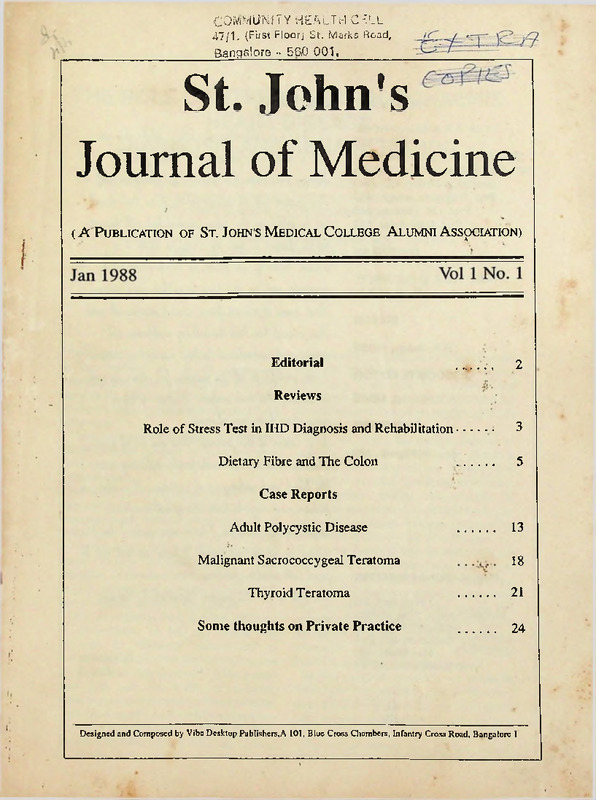
- Title
- ST. JMC JOURNAL OF MEDICINE ISSN 0970-4221 VOL.I NO. 1 JANUARY 1988
- extracted text
-
community health cell
47/1. (First Floor; St. Marks Road,
Bangalore - 5&0 001,
St. John's
Journal of Medicine
(A Publication of St. John's Medical College Alumni Association)
Editorial
. _ . ,
2
Role of Stress Test in IHD Diagnosis and Rehabilitation.........
3
Reviews
Dietary Fibre and The Colon
■...........
5
...........
13
Case Reports
Adult Polycystic Disease
18
Malignant Sacrococcygeal Teratoma
Thyroid Teratoma
...........
21
Some thoughts on Private Practice
...........
24
Designed and Composed by Viba Desktop Publishers,A 101, Blue Cross Chambers, Infantry Cross Road, Bangalore 1
PATRONS
Prof. A.F.A. Mascarenhas,
MS, FRCS
Prof. (Lt. Gen.) G.R. Narayanan,
MD, DM, FAMS, FACC, F. LAMAS
Prof. P.S. Shetty, MD, PhD
Prof. Thangam Joseph, MD
Prof. I.M. Thomas, MS
Prof. Dara Amar, MD
OVERSEAS
Salim Yusuf, D.Phil, MRCP,
N.I.H (U.S.A)
EDITORIAL
H. fydvn\ ib odd^ali/ny, iIa, iilwA, ^JuIm, Uua, uhjoa,
a/nd a fu
Alumni, Av^ciali&n Im tcJwn, Uua, laJv up&n, ilAel^.
JkiA j&uA/rml unit Im, umujua i/n, (La M/nM, dud iL axil
on-
oLmtmw all wn, (La uxmld
ujjccjiaa.
3ndiallip uh, i/nlwd (a-
iL vid, oa, a,
(M/AAluJy, a/nd uh, aAo k&pA io pulliAh pw\, 'wxjuua
oLguL iAAM of, i/rd/wd
EDITOR
makb iL a,
upi/Ltd uipvJA, all gwa,.
JIua, j&u/vnal Im had ilb lullwn^ pAjMi/M] nid
R.R. Baliga, MBBS
ma/ni^ p&ipts, wduniM/vid io hdp duib io W«/uxiito*nA..
ASSOCIATE EDITOR
3 oati ^AaLtlul io all tfvb pal/ioM 1^, Uvua, Mipp^d,
A.V. Kurpad, MBBS
PUBLISHER
i/n, doAti/n^ I/ua, mw- (H/nluAtb. 3 i/ipuL a, Id 9^
CMp9AoLiA4i,
oo/v alwrTvru,.
/ta 0/ Sdila^ d UiiA, juwnjal, Uvm oaa Id d iAAM}
Arun B. Kilpadi, MS
udiick ha/H, io In, JaoIL unlk [iA/mly, unduddny, (aam
biaud amd cadii^, up
daliAlkA,. 31 ia> oIao a,
loAhu^Tb n9u>ada^A, io hoM, ma/ruf auUuMA'
q,
papuA,, ^TidurM 3 hoM, mumv ty> io 7 io 9
PUBLICATIONS COMMITTEE
Prem Pals, MD
Prakash Appaya, DPM, MNAMS
Ranganath Nayak, MD
S.C. Rajendran, MD, DVD
M.H. Sheriff, MD
Kevin Mascarenhas, MBBS
We, hope,
yd Uua, joufi/nal wyiAitAjtd
Uvb rwd
iAAUJb.
R.R.Baliga
MBBS
THE ROLE OF STRESS TEST IN ISCHAEMIC
HEART DISEASE
DIAGNOSIS AND REHABILITATION
LT. GENERAL G.R. NARAYANAN MD., DM (Cardiology), FACC, FAMS, FIAI
PROFESSOR AND HEAD, DEPT. OF CARDIOLOGY
ST. JOHN'S MEDICAL COLLEGE AND HOSPITAL
BASED ON THE ORATION DELIVERED IN MEMORY OF LATE
DR. JAYASHREE THOMAS -30.11.87
This paper is based on Oration, dedicated
to the memory of late Dr. Jayashree
Thomas, and related mainly on the work
done at the Dept, of Cardiology, St. John’s
Medical College Hospital from January 1986
till to-date.
The latter part - rehabilitation of Ischaemic
Heart Disease patients, is from the work
done at the Armed Forces medical Services
establishment.
Ischaemic Heart Disease (coronary artery
disease) has a very broad spectrum in its
manifestations. While at one extreme it may
remain silent or asymptomatic; on the other,
it may come as a ’bolt' from the blue with an
acute severe episode of myocardial
infarction. In between are the chronic stable
angina, variant and unstable angina, ECG
abnormalities, rhythm disturbances.
The role of Stress Test is in the detection
or confirmation of IHD, its extent and
severity, its follow-up, assessment of
therapy, future direction for further invasive
investigations like Coronary Angiography
and last but not least, in planning
rehabilitation programme for these patients.
We have used CASE - II Marquette Elec
tronics Computerised Treadmill System.
This has facilities for simultaneous 12 Lead
analysis which are displayed throughout the
test with arrhthmia leads for continuous
monitoring, Heart rate, BP response and
symptoms display, and most important the
Computerised ST segment response and its
scope.
When we stress the cardiac muscle by
exercise, the myocardial oxygen demands
goes up depending on the severity and
duration of exercise. In normal individuals,
the coronary arteries are capable of
supplying the demand. In diseased vessels,
this does not happen and hence myocardial
ischaemia is brought out or enhanced.
Using the Treadmill test, the end points are
significant ST depression (more than 1 mm.
horizontal, downsloping or slow upsloping)
symptoms of angina or dyspnoea, the heart
rate and BP response. Other events like
arrhythmias are carefully watched for. It is
mandatory to have cardiac resuscitatory
measures, including DC defibrillator
available in the Stress Lab.
Various exercise protocols are available
suitable for the individual patient. We have
used BRUCE PROTOCOL for most of the
patients with modification in a selected few
early stress tests.
The test is carried on until the subject
4
ST. JOHN’S JOURNAL OF MEDICINE
achieves his predicted maximal heart rate
(based on age and sex) or on occurence of
symptoms or significant ST changes or
serious arrhythmias.
We also observe the changes during
recovery phase for about ten minutes.
Based on the above, we have performed
more than 500 Stress Tests during this
period without any untoward event. They
have been performed in cases of Angina, a
typical chest pain, ECG abnormalities, Post
Myocardial
Infarction,
Arrhythmias,
controlled hypertensives and routine
executive cardiovascular check-up.
In cases of Angina, stress test has been
useful in assessing the severity of the
underlying coronary artery disease and
future direction of therapy and its efficacy as
well as for coronary artery by-pass surgery
(CABG) or angioplasty as indicated. Where
we found significant ST depression in the
early part of the exercise, we have advised
early coronary angiogram, there patients
have shown triple vessel disease or left main
involvement. In atypical chest pain, stress
test has been useful in detecting ischaemic
heart disease while in a significant group it
has been useful in reassuring the subject
that the chest pain is not cardiac in origin.
The same applies to those with non-specific
ECG abnormalities, some of whom come
under the group of vasoregulatory
abnormality.
We have carried out stress test serially in
Post Mysocardial Infarction cases. The test
has been useful in assessing the follow-up,
improvement or deterioration and for
evaluation for CABG. We have also carried
out early Stress test (within 6 weeks) in a few
cases of Mycocardial Infarction treated with
IV Streptokinase, as well as post CABG and
post angioplasty. Stress test after post
Myocardial Infarction indicates good
prognosis, where there is no fresh
ischaemia; they can be rehabilitated with
significant presence of ischaemia. Will early
coronary angiogram for assessment of
CABG or angioplasty. In IV streptokinase
Vol. 1 no. 1
less than 10% required angioplasty.
Assessment of arrhythmias associated
with IHD , their unmasking and effects of
therapy is another facet of stress test.
It is useful in detecting asymptomatic or
silent ischaemis as has been our experience
in the Armed Forces as well as at this
Institution when we performed stress test on
executives above fifty years age as a
rountine CVS check-up.
Our experience in the Armed Forces points
to the great value of stress test in
rehabilitation of Ischaemic heart Disease
patients by graduated exercise programmes
in addition to other measures. Most of the
patients could be sent for fruitful
continuation of their employment in the
Armed forces and some for even very
strenuous physical work including return to
battle-field and adverse environmental
conditions like high altitude.
However, there are a few pitfalls and
limitations in Stress test including false
positive and false negative, as in W-P-W
syndrome, Mitral value prolapse, bundle
branch block and cardiomyopathies.
REFERENCES
DeBUSK (R F) et al.
Submaximal Predischarge exercise testing
after acute myocardial infraction: who needs
It?
Am J Cardiol 55(4):499-500 Feb.1985
2. KRONE (R J), et al.
Low-level exercise testing after myocardial
infarction: usefulness In enhancing clinical
risk stratification.
Circulation 71(1):80-9 Jan. 1985
3. MUKHARJI (J), et al.
Right positive exercise test and extensive
Coronary disease: effect of antlanginal
therapy
Am J Cardiol 55(4):277-80 Feb.1, 1985
4. WIJNS (W) et al.
Predictive value of early maximal exercise
test and thallium scintigraphy after successful
percutaneous tranlumlnal coronary
angioplasty
Br. Heart J 53 (2):194-200 Feb.1985
1.
DIETARY FIBRE AND THE COLON
A.V. KURPAD, MBBS
P.S. SHETTY, MD.,PhD
DEPARTMENT OF PHYSIOLOGY
Dietary fibre is a comparitively recent
phenomenon. A hitherto indifferent
response towards the beneficial effects of
this substance by the medical fraternity has
been shaken up mainly by the effort of
Denis Burkitt, whose seminal monograph'
in 1975 marked an explosion of interest in
this field. Subsequently, every property of
fibre has been extensively studied, and
though fibre is known to affect gastric
intestinal function extensively, far ranging
effects on blood clotting factors,2 sex
hormones3 and so on have also been
examined. Evidently, there is more to fibre
than meets the eye. In retrospect, it is
instructive to note that ancient India and
Greece recognised the beneficial effects of
unrefined food. The Charaka Samhita
(without a reference to which no review is
complete!) also ascribes obesity and
diabetes to refined cane sugar.
What is Dietary fibre?
Dietary fibre is defined as a group of
polysaccharides associated with the cell
wall of plants, which are resistant to the
digestive effect of endogenous secretions
of the human gut.9 While this definition is
both chemical as well as functional; it is also
incomplete, as some constituents of die
tary fibre are neither polysaccharides, nor
are they cell wall associated,*-7 and some
starch and protein also escape digestion in
the gut.*-® In addition fibre invokes intuitive
images of something fibrous’, which is
unfortunate; many components of the
defined dietary fibre are gums and
mucilages-which are certainly not fibrous.
In sum then, there is some confusion as
to exactly what fibre is, or more specifically,
what it should be. Broadly, one could simply
say that dietary fibre is of plant origin and
that it is relatively resistant to digestion.
Dietary fibre is a phenomenon; to attempt
to review it completely is fanciful. This
review confines itself to the actions of fibre
on some aspects of colonic function. Other
actions of dietary fibre are excellently
reviewed elsewhere.”-”
Among the effects of fibre, the one which
is most evident, is it's effect on faecal
weights. Faecal weights of populations are
known to vary depending on the
geographical location, diets and a host of
other factors. Faecal weights range from
about 125g/day12-13 in the West to about
300g/day in the Tropics.'4 The faecal
weights of an individual can vary from 19
to 1500g/day.'4 While the range of faecal
weights is well known, at least one
important fact is in doubt. That is, what are
the mechanisms of faecal bulking in man
and how does dietary fibre contribute to it?
Since faeces was considered to be
composed primarily of dietary residue, the
search began for principle dietary
constitutents which could affect faecal
output. Controlled studies have shown that
6
ST. JOHN'S JOURNAL OF MEDICINE
changing fat and protein intake do not affect
faecal weight”-’8'17 Dietary carbohydrate,
however, in the form of starch showed an
increased faecal bulk with increased
intakes.18 There has also been an explosion
of knowledge about the alternate form of
carbohydrate in diet, (i.e) dietary fibre. A
very strong correlation has been drawn
between fibre and faecal weight.18'87 Further
investigation has shown that different types
of fibre bulk faeces to different extents.80
Many theories have been invoked to explain
the bulking action of fibre including its
physical characteristics, its chemical com
position (especially pentoses) and its role
as a substrate for bacteria.21
The initial mechanism ascribed to fibre
invoked its water holding capacity.22 Since,
in addition, it was by then well known that
fibre resisted digestion, it was thought that
this resistant fraction of the dietary intake
absorbed water, swelled and increased in
size, and hence, bulked the faeces. This
view was strengthened by the fact that
there was a significant relationship between
the initial transit time (ie.) the fastest and
the faecal bulking achieved.20-32
In contrast however, it was also shown
that there was an inversely linear relation
ship between the invitro water holding
capacity of fibre and the effects the same
fibre had on colonic function.23 Evidence
was also presented against the apparent
indigestibility of fibre. Studies by Williams
and Olsted in 1936 and by Stephen showed
that the degree of digestibility of fibre varied
with other factors like the type of fibre as
well as the transit.24-28
In addition a recent assessment of
microbial content of human faeces showed
that bacterial represented a high proportion
of faecal mass.28 Since half the faecal bulk
on a British diet was bacteria (this study
was from a British group), alterations in
colonic flora provided ample scope for
altering faecal weight This led to the
postulation that many types of fibre act as
substrates for colonic flora, and the
Vol. 1 no. 1
resultant increase in microbial growth bulks
the faeces.28 Anaerobes, which are a major
constitutent of colonic microbes are known
to digest fibre. The anaerobic digestion of
fibre, called fermentation, yields 3 products;
SCFA’s (Short chain fatty acids) gas and
energy.27,44 The growth of colonic bacteria
is accomplished by use of this energy and
consequent fixation of Nitrogen in the gut.
Major limitations of this theory included the
amount of time needed to metabolize
fibre28 as well as the adequacy of substrate
available.30 It is evident that, in fast transit
states at least, there must be very little time
for fermentation of fibre.28'28
To support the role of adequate substrates
for bacteria, came the concept of the
‘carbohydrate gap'.30 It has been estimated
that supporting a population of bacteria in
the colon would require 70 g of carbo
hydrate per day. In India dietary fibre
intakes range from 20-30 g/day.31 The
remainder 40-50 g carbohydrate require
ment is termed the ‘carbohydrate gap’ and
it is postulated that undigested starch
(which could be upto 10-20%) fills this gap.
In India, starch intakes are high, and this
could account for the high faecal weights
seen in the tropics, despite relatively low
intakes of fibre.32
Thus, two clearly defined mechanisms
exist, to explain the mechanism of faecal
bulking. In the light of the high faecal
weights and low transit times seen in the
tropics, it is important to elucidate the
dominant mechanism. The water holding
hypothesis has been tested by inhibiting the
normal fermentative process. This was
done by administering Metronidazole
orally,28 to a group of human subjects.
Metronidazole, which is particularly
effective against anaerobes, decreased
fibre degradation significantly and
increased faecal weight. While this
substantiates the fact that a large fraction
of dietary fibre is digested by the colonic
flora, it also suggests that water holding by
undigested fibre is a far more effective
JAN. 1988
ST. JOHN’S JOURNAL OF MEDICINE
mechanism for faecal bulking. In fact, it
follows that any degradation of fibre would
relatively decrease faecal bulk, since, for
the same amount of fibre, faecal weights
are much larger if colonic flora are
prevented from digesting the fibre.
On the other hand, bacterial mass is not
to be ignored. In a study where starch
intake was increased,33 it was seen that
faecal weights also increased, though not to
the extent seen when fermentation was
inhibited by metronidazole. However, the
increase in faecal weight in this instance
was due to an increased bacterial mass, as
evinced by the increase in faecal weight in
this instance was due to an increased
bacterial mass, as evinced by the increase
in faecal Nitrogen. It is clear that, given
adequate substrate, bacterial mass has a
role to play in faecal bulking, however, vis*
a-vis fibre, its role is doubtful.
Another important effect of fibre is on
intestinal transit time. Much has been said
on the subject, and the essential conclusion
is that dietary fibre reduces transit time.34
The mechanism invoked is one of a greater
faecal mass which stiumlates motility and
therefore reduces transit time. This simple
and attractive picture presents problems.
For one, the inverse relationship between
faecal weight and transit time suggests that
a simple cause and effect relationship may
not exist. Could not, for instance transit time
be the dominant determinant variable, and
therefore faecal weight be the dependent
subordinate process? Transit time maybe
important in determining many reactions in
the colon, for example, the breakdown of
salicylazo sulphapyridine (SASP) in the
gut.35 Secondly, the inverse relationship
cited is curvilinear; thus a point exists at the
asymptote, the so called critical faecal
weight (which is about 150g) beyond which,
any further increase in faecal weight has no
associated transit time change. Ethnically,
data points on the linear part of this curve
are all derived from British subjects, while
Indians lie on the asymptote. In terms of
7
transit time therefore, one must be careful
when describing the effect of fibre. While it
will certainly bulk the faeces, there may be
no attendent change in transit.
Another problem with transit is the site.
Upto now, the term transit, when used,
implied total mouth to anus transit time.
However, the dominant effect of fibre is to
speed up colonic transit: in fact, fibre
actually slows transit through the stomach
and small intestine.36-3738 One study has
claimed that fibre actually alters transit to
a salubrious (utopian? as sufferers of
constipation or IBS would feelingly say) via
media.29 That is, fibre shortens transits
where transits are long, and lengthens
transits when they are short. These glad
tidings were not confirmed however; fibre
still remains most effective in the 'sensitive
population’, previously described. Interes
ting offshoots exist. One example: a recent
study which demonstrated a reduction of
transit time in pigs fed indigestible grains of
polyethylene which resembled rice.40 Has
the age of plastic finally caught up with what
we eat?
The breakdown of fibre in the large
intestine is not without its attendant
consequences. As mentioned earlier, fibre
fermentation gives rise to SCFA’s, energy
and gas.41 This last is possibly the most
serious 'social' side effect of fibre, and
probably points to why people moved away
from unrefined foods earlier in this century.
The gases generated are Hydrogen and
Methane and contribute to flatus and foul
breath.42 Knowing the existence of gases
makes the phenomenon of a colonic
explosion during polypectomies with poor
preparation vey real.43 As said earlier, the
only other damages are of a social nature;
for who has not been unwise in the eating
of dal or beans and has not later repented?
The major SCFA’s are acetates,
propionates and butyrates. They are
available for absorption, and hence could
be considered a putative energy source
(albeit almost negligible) not only in the
8
ST. JOHN’S JOURNAL OF MEDICINE
body,44 but also for the enterocyte,45 and
their absorption is attended by increased
sodium and water absorption.46 Osmotic
effects of unabsorbed SCFA’s cannot be
ignored, even though they are generated in
small amounts and importantly, butyrates
have been shown to have an inhibitory
effect on anaplastic growth47 and a hyper
proliferative effect on colonic epithelium.48
Applications of fibre
As far as the colon is concerned, the
protective effect of fibre has been
extensively studied with regard to colorectal
cancers as well as constipation. There is a
strong suggestion that fibre deficient diets
are associated with colorectal
carcinoma.4® The protective effect of fibre
has been amply demonstrated in Africa;
yet, two questions need to be addressed.
One, is the protective effect really extant,
and two, how does it work? Some detailed
studies of colonic cancer have shown that
there is a weak protective effect of fibre.50-51
However, given the multiple aetiology of
colonic cancer, some studies have indeed
found the protective effect of fibre to be
negligible.52-53 Animal models are also
equivocal; while some demonstrate
protective effects, others do not.54-55-56
Indeed, one study has also shown a higher
incidence of tumours when wheat bran was
used.57 The popular notion however, is that
fibre still protects. The mechanism of this
protection may be by dilution of carcinogen
by water holding, or by reduction of transit,
and hence exposure time. Faecal ammonia
falls58 (probably due to N fixation by growing
bacteria) as do bile acid concentrations.58
The dehydroxylation of bile acids is
prevented by fall of pH due to the
generation of SCFA’s.60 This reduces the
concentration of deoxycholate, while
increasing chenodeoxycholate: a salutary
effect.61 Carcinogens, like bile salts may
also be bound and made ineffective.62
SCFA’s have their own inhibitory effect on
Vol. 1 no. 1
malignant change.47 Fibre also reduces
mucin degradation by bacteria in rats.63
Conversely fibre fermenation (with
cellulose) reduces faecal bacterial Bgalactosidase and B-acetylglucosaminidase which preserve the integrity of
the colonic mucosa.64 Thus the facts are
many, and diverse; many of them are
experimental or in vitro findings — to extra
polate them to an in vivo situation could be
dangerous. It needs to be seen which
factors and mechanisms are dominant and
important.
With regard to constipation, fibre has its
best known effect; certainly, a fibre
depleted diet is associated with cons
tipation and attendant sequelae.65-66 It is
likely that this problem is the root cause of
the enormous research in dietary fibre;
indeed, the following lines aptly sum up the
angst of the constipated individual:
There has never lived a poet in the whole
history of the world, ancient or modern, near or
far, who ever managed to write great poetry, or
even passably fair and decent poetry, at a time
when he was suffering from stenosis at any
point along the 34 foot Via Dolorosa running
from the pylorus to the sigmoid flexure.... He
is stumped and helpless. The more he tries, the
more vividly he will be conscious of his
impotence. Sweat will stand out in beads upon
his brow, he will fish patiently for the elusive
thought, he will try coaxing and subterfuge, he
will retire to his ivory tower, he will tempt the
invisible powers with black coffee, tea, alcohol
and the alkaloids-but he will not write his poem,
or Iron out his syllogism, or get his fleshstone
or perfect his swindle; striving In the face of such
an interior obstacle Is the most cruel of
enterprises.... *
A Mencren Chrestomathy
-H.L Mencken (1949)
Diverticulosis has been attributed to high
intracolonic pressure and is charateristic of
the West.67 Fibre is said to help in such
cases, though there are misgivings with this
hypothesis68 IBS too, has a fibre clique
which believes that the problem of
JAN. 1988
ST. JOHN’S JOURNAL OF MEDICINE
disordered motility is secondary to the
faecal bulk. Some studies have indeed
shown that fibre does have an alleviating
effect on the problem.®070-71
Important extra alimentary applications of
dietary fibre are diabetes mellitus,
cholesterol gallstones, obesity, athero
sclerosis and ischaemic heart disease.
Fibre depleted diets are epidemiologically
associated with diabetes,72 and it is possible
to experimentally create diabetic animal
models with fibre depletion.72 The
mechanism of protection has been
hypothesized to be the attenuation of post
prandial hyperglycaemia73-74 with fibre;
repeated hyperglycaemic peaks are known
to be associated with insulin resistance.75 In
fact, it is entirely possible that fibre may
help diabetic individuals by balancing the
glycaemic challenge and the insulin
response evoked. Gallstones are a
common western phenomenon7® and fibre
has been shown to experimentally reduce
the lithogenic potential of bile.77
Mechanisms here invoke the binding of bile
salts and consequently diminished re
absorption78 which is beneficial, since
chenodeoxycholate reduces cholesterol
synthesis.80 There are at least three mecha
nisms by which fibre prevents obesity. It
displaces nutrients (by dilution; slows food
intake and causes satiety (by distending the
stomach), and reduces the digestibility of
nutrients.81 Fibre seems to have a
protective effect on atherosclerosis and
IHD82 and affects lipoprotein fractions,83 as
well as lowers serum cholesterol.84
In conclusion, it appears that fibre is good.
Arguments to the contrary (not that fibre is
bad, but that no fibre may also be good)
quote the Masai and Eskimaux tribes who
takes almost no food of vegetable origin.85
The case against this rests on obvious
grounds, however, it is clear that fibre has
not yet given up all its secrets. Let us
simply, for the present, state that fibre is
necessary, though how much is needed is
still in dispute. Based on ideal faeal weights
9
and ideal transits, a proposal has been
made that at least 30g of fibre should be
taken per day.86 Indian intakes (on which we
do very well ) are somewhere around or
below this mark, though starch probably
plays a bigger role.31 The sources of fibre
should be natural, preferably cereal based,
the case for this cannot be
understated.10-87M
Earlier investigators had to work uphill on
popularising fibre. Their refrain was “It may be
s..t to you, but it's bread and butter to usl".
Clearly fibre is bread and butter for all of us and
it is wise to heed the famous words of Ogden
Nash: “There is nothing so overrated as the
orgasm, and nothing quite so underated as a
good stool I".
REFERENCES
1. Burkitt DP. Trowell HC, eds. Refined
carbohydrate foods and disease. Some
Implications of Dietary Fibre. London:
Academic Press. 1975.
2. Simpson HCR, Manu JI Chakrabarti R et al.
Effect of high fibre diet on haemostatic
variables in diabetes. Br. Med J 1982:284:
1608
3 Hamlalnen EK, Aldercreutz H. Puska P,
Pietinen P Decrease of serum total and free
testosterone during a low fat high fibre diet.
J. Steroid Blochem 1983, IS 369
4. Me Cance RA, Widdowson EM. Old
thoughts and new work on breads white and
brown. Lancet 1955:i:205
5. Trowell H. Southgate DAT. Wolever TMS,
Leeds AR, Gassul MA, Jenkins DJA. Dietary
fibre redefined. Lancet 1976:1:967
6. Cummings JH. What is fibre? In: Spiller GA.
Amen RJ eds. Fibre in human nutrition. New
York: Plenum Press, 1976: 1-30.
8. Hellendoorn EW. Dietary fibre or
indigestable residue ? Am J Clin Nutr 1981;
34; 1437
9. Saunders RM, Betchart AA The signif
icance of protein as a component of dietary
fibre. Am J Clin Nutr 1980;33; 960-961
10. BIJIanI RL. Dietary Fibre: consensus and
controversy. Progress In food and nutrition
science 1985; 9:343.
11. Vahouny GV, Kritchevsky D, eds. Dietary
Fibre In health and disease. New York:
10
ST. JOHN’S JOURNAL OF MEDICINE
Plenum Press, 1982
12. Wiggins HS, Cummings JH, Evidence for
mixing of residue In the human gut. Gut
1976, 17:1007.
13. Wyman JB. Heaton KW, Maning AP, Wicks
ACB. Variability of colonic function in healthy
subjects. Gut. 1978;19:146
14. Cummings JH. The colon. In: Bouchier IAD
ed. Recent Advances in Gatroenterology.
London: Churchill Livingstone, 1980:87
15. Cummings JH. Wiggins HS. Jenkins DJA,
Houston H, JlvraJ T , Draser BS. Hill MJ.
Influence of diets high and low in animal fat
on bowel habit, gastrointestinal transit time,
fascal microflora, bile acid and fat excretion
J Clin Inv. 1978: 61:953.
16. Calloway DH. Kretsch JM. Protein and
energy utilization In men given a rural
Guatemalan diet and egg formulas with and
without added oat bran. Am J Clin Nutr
1978L31:118
17. Cummings JH. Hill MJ, JivraJ T, Houston H.
Branch WJ Jenkins DJA. The effect of meat
protein and dietary fibre on colonic function
and metabolism part I Changes is bowel
habit, bile add excretion and calcium
absorption Am J Clin Nutr 1979, 32:2094
18. Shetty PS. Studies in Protein and Energy
restriction and dietary thermogenesis in
obesity and chronic undemutrition, PhD
Thesis 1980, University of Cambridge.
19. Cummings JH. Physiological aspects of
dietary fibre In man. In. Truelove SC.
Mayworth MF. ed.s Topics In Gastro
enterology, Oxford, Blackwell 1987:49.
20. Cummings JH. Southgate DAT, Branch WJ,
Houston H, Jenkins DJA, James WPT,
colonic response to dietary fibre from carrot,
cabbage, apple, bran and guar gum: Lancet
1978; l:5
21. Cummings JH. Consequences of the
metabolism of fibre In the human large
intestine. In: Vehouny CV, Kritchevsky
D.Eds, Dietary fibre in health and disease.
New York: Plenum Press, 1982:9.
22. Eastwood MA. Dietary fibre in human
nutrition. J Sd Food Agr 1974; 25: 1523
23. Stephen AM, Cummings JH. Water holding
by dietary fibre In vitro and its relationship
to faecal output In man. Gut 1979; 20: 722
24. Williams RD, Olmsted WH. The manner In
which food controls the bulk of the faeces.
Ann Int Med 1936;10:717
Vol. 1 no. 1
25. Stephen AM, Cummings JH. The microbial
contribution to human faecal mass. J Med
Microbiol 1980;13:45
26. Stephem AM, Cummings JH. Mechanism of
action of dietary fibre in the human colon.
Nature 1980;284:283
27. Caspary WF, Lembcke B. Elsehans B.
Bacterial fermentation of carbohydrates
within the gestro intestinal tract. Clin Res
Rev 1981; 1: 107
28. Kurpad AV. Shetty PS. Effects of anti
microbial therapy on faecal bulking. Gut
1986;27:55
29. Stephen AM. Dietary fibre and human
colonic function. PhD Thesis 1980,
University of Cambridge.
30. Stephen AM. Haddad AC, Philips SF.
Passage of carbohydrate into the colon.
Direct measurements in humans.
Gastroenterology 1983; 85:589
31. Shetty PS, Kurpad AV. Intestinal transit
times of S. Indian subject. Ind. J Med Res
1984;80:693
32. Gopalan C,
Ramasastri BV, . Balasubramanilam SC. Nutritive value of Indian
Foods New DelhkICMR 1980.
33. Shetty PS. Kurpad AV. Carbohydrate Intake
influences faecal bulking. Am J Clin Nutr
1985;43:210.
34. Hillman L, Peters S, Rsher A, Pomare EW.
Differing effects of pectin, cellulose and
ligmin on stool pH. transit time and faecal
weight. Br. J.Nutr 1983, 50:189
35. Van Hees PAM, Tulnto JHM, Van Possum
Jm, Van Tongeren JHM. Influence of
intestinal transit time on azo reduction of
Salazopyrin. Gut 1979; 20:300
36. Me Cance RA, Prior kM, Wlddowson EM. A
radiological study of the rate of passage of
brown and white bread through the digestive
tract of man. Br J Nutr 1953; 7:98
37. Grimes DS. Goddard J. Gastric emptying of
whitemeal and white bread. Gut 1977;
18;725
38. Fleramonti J, Bueno L, Ruckebusch Y.
Dietary fibre an motor profile of small
intensive and hindgut. In: Wallace G. Bell L.
eds. Fibre in Human and animal nutrition.
Wellington: the Royal Society of New
Zealand, 1983:59
39. Harvey RF, Pomare EW, Heaton KW.
Effects of Increased dietary fibre on
intestinal transit. Lancet 1973; 1:1278
JAN. 1988
ST. JOHN’S JOURNAL OF MEDICINE
40. Cherbut C. Ruckebusch Y. The effect of
Indigestible particles on digestive transit
time and colonic motility In dogs and pigs Br
J Nutr 1985; 53:549
41. Cummings JH. Short Chain fatty acids in the
human colon. Gut 1981;22:763
42. Marthlnsen D, Fleming SE. Excretion of
breath and flatus gases by humans
consuming high fibre diets. J Nutr 1982;
112:1133
43. Bigard MA, Gaucher P. Las al I e C. Fatal
colonic explosion during colonoscopic
polypectomy. Gastroenterology 1979;
77:1307
44. Me Nell Nl, Cummings JH, James WPT.
Short chain fatty acid absorption by the
human large intestine. Gut 1978; 19:819
45. Roediger WE. Utilization of nutrients by
isolated epithelial cells of the rat colon.
Gastroenterology 1982; 83:424
46. Roediger WE, Moore A. Effect of short chain
fatty acid on sodium absorption In Isolated
human colon perfused through the vascular
bed. Dig Dis Sci 1981 ;26:100
47. Hagoplan HK. Riggs MG. Swartrz LA. Effect
of m butyrate on DNA synthesis In chick
fibroblasts and hela cells. Cell 1977;12:855
48. Jacobs LR. Lupton JR. Effect of dietary
fibres on rat large bowel mucosal growth
and cell proliferation. Am J Physiol
1984,246:4378
49. Burkitt DP. Etiology and prevention of
colerectal cancer. Hosp Pract (off) 1984;
19:67
50. Cummings JH. Epidemiology of fibre Intake
and disease. In: Wallace G. Bell L, eds.
Fibre In human and animal nutrition.
Wellington: The Royal Society of new
Zealand, 1983:11
51. Graham S, Dayal H. Swanson M, Mittleman
A, Wilkinson G. Diet In the epidemiology of
cancer of the colon and rectum. J Nut
Cancer Inst. 1978; 61:709
52. Armstrong B. Doll R. Environmental factors
and cancer Incidence and mortlalty In
different countries with special reference to
dietary practices. Int. J Cancer 1975; 15:
617-631
53. Lin K, Stamler D. Moss D. Garside D,
Persky V, Soltero I. Dietary cholesterol, fat
and fibre, and colon cancer mortality. Lancet
1978; ll:782
54. Freeman JH, Spiller GA, Kim YS. A double
II
blind study on the effects of differing purified
cellulose and pectlne fibre diets on 1,2
dimethyl hydrazine Induced rat colonic
neoplasia. Cancer Res 1980;40:2661.
55. Reddy BS. Mori H. Nicolais M. Effect of
dietary wheat bran and dehydrated citrus
fibre on azoxymethane Induced intestinal
carcinogenesis In Fischer 344 rats. J Nut
Cancer Inst. 1981; 66:553
56. Cruse JP, Lewin MR. Clark CG. Failure of
bran to protect against experimental colon
cancer in rats. Lancet 1979; II: 1278
57. Jacobs LR. Enhancement of rat colon
carcinogenesis by wheat during the stage of
1,2 dimethylhydrazine administration.
Cancer Res 1983;43:4057
58. Cummings JH, Branch WJ. Postulated
mechanisms whereby fibre may protect
against large bowel cancer. ln:Vahouny GV.
Kritchevsky D. eds. Dietary fibre in health
and disease. New York: Plenum Press.
1982:313
59. Cummings JH, Hill MJ, Bone ES, Branch
WJ, Jenkins DJA. The effect of meat protein
and dietary fibre on colonic function and
metabolism. 11’.Bacterial metabolltles in
faeces and urine. Am J Clin Nutr 1979;
32:495
60. Walker ARF, Walker BF, Segal I. Faecal pH
value and Its modification by dietary means
in South African black and white school
children. A Med J 1979;55:1081
61. Thornton JR. High colonic pH promtoes
colorectal cancer. Lancet 1981; 1:1081.
62. Smith - Bonbaro P. Hanson D, Reddy B.
Carcinogen binding to various types of
dietary fibre. J Nut Cancer Inst. 1981;
67:495
63. Shlan S, Chang G. Effects of dietary fibre on
faecal muclnase and geucuronldase activity
In rats. J Nutr 1983; 113.138
64. Prizent DP. Influence of high dietary
cellulose on fecal glycosidases In
experimental rat colon carcinogenesis.
Cancer Res 1984;44:557
65. Burkitt DP. Walker ARP, Painter NS. Effect
of dietary fibre on stools and transit times,
and Its role In the causation of disease.
Lancet 1972;ii:1408
66. Cummings JH. Constipation, dietary fibre
and the control of large bowel function. Post
grad med J 1984;60:811.
67. Painter NS. Diverticular disease of the
12
ST. JOHN’S JOURNAL OF MEDICINE
colon. Br Med J 1968;3:475
68. pastwood MA, Smith AN, Brydon WG.
Pritchard J. Comparison of bran, ispaghula
and lactulose on colon function in
diverticular disease. Gut 1978; 19:1144
69. Cann PA, Read NW, Holdworth Cd. What Is
the benefit of coarse wheat bran in patients
with Irritable bowel syndrome? Gut 1984;
25:168
70. Golecha AC, Chadda VS. Chadda S,
Sharma SK, Mishra SN. Role of ispaghula
husk In the management of Irritable bowel
syndrome: A randomized double blind cross
over study. J Ass Phy Ind 1982; 30:353
71. Kumar A, Kumar N, VIJ JC, Sarin SK. Anand
BS. Optlnum dosage of Ispaghula husk in
patients with Irritable bowel syndrome:
correlation of symptom relief with whole gut
transit time and stool weight. Gut 1987;
28:150
72. Trowell H. Diabetes Mellitus and obesity. In
Burkitt DP. Trowell HC, eds. Refined
carbohydrate Foods and Disease. Some
Implications of dietary fibre. London:
Academic press, 1975:227.
73. Williams DRR. James WPT. Fibre and dia
betes. Lancet 1979:1:271
74. Monnler LN. Lotman MJ, Collette C.
Monnler MP, Mlrouze J. Effects of dietary
fibre supplementation in stable and labile
Insulin dependent diabetics. Dlabetologla
1981; 20:12.
75. Wright DW, Hansen Rl. Mondon CE.
Reaven GM. Sucrose induced insulin
resistance in the rat modulation by exercise
and diet. Am J Clin Nut. 1983; 38:879
76. Heaton KW, Gallstones and cholecystitis.
In: Burkitt DP. Trowell HC eds. Refined
carbohydrate Foods and disease. Some
Implications of dietary fibre. London:
Academic press. 1975:173
77. Kritchevsky D, Tepper SA, Klurfeld DM.
Effect of pectin and cellulose on formation
and regression of gallstones In hamsters.
Experlentia 1984; 40:250
78. East wood MA, Hamilton D. Studies on the
absorption of bile salts to nonabsorbed
components of the diet. Blochim Blophys
Acta 1968; 152:165
79. Pomare EW, Heston KW, Low Beer TS.
Esplna HJ. The effect of wheat bran upon
bile salt metabolism and upon the lipid
composition of the bile In gallstone patients.
Vol. 1 no. 1
Am J Dig Dis 1976; 21:521
80. Coyne MJ, Bonarris GG, Goldcstein LI,
Shoenfield IJ. Effect of chenodeoxy cholic
acid and phenobarbital on the rate limiting
enzymes of hepatic cholesterol and bile acid
synthesis in patients with gallstones. J Lab
clln med 1976;87:281
81. Heaton KW. Food fibre as an obstacle to
energy intake. Lancet 1973; 11:1418
82. Morris JN, Marr JW, Clayton D. Diet and
heart: a postscript. Br Med J 1977;2:1307
83. Kirby RW, Anderson JW, Sieling B. Rees
Ed, Chen WL, Miller RE, kay RM. Oat bean
intake selectively lowers serum low density
lipoprotein cholesterol concentrations of
hypercholesterolaemic men. Am J Clin Nutr
1983; 38:285
84. Bijlani RL, Gandhi BM, Gupta MC, Manocha
S., Tandon BN. Effect of whole buckwheat
flour supplementation on lipid profile and
glucose tolerance. Ind J Med Res
1985;81:162.
85. Eastwood MA, Passmore R. Dietary fibre.
Lancet 1983; ii:202
86. Mann JI. Diseases of overnourished
societies and the need for dietary change.
ln:Weatherall DJ. Ledlnghan JGG, Warrell.
eds. Oxford Textbook of Medicine. Oxford:
Oxford University Press. 1983:55.
87. Trowell H. Ischaemic heart disease,
atheroma and fibrinolysis. In: Burkitt DP,
Trowell HC eds. Refined Carbohydrate
Foods and disease. Some Implications of
Dietary Fibre. London: Acadamic Press,
1975:195
88. Kay RM Truswell S. Effect of citruspectin on
blood lipids and faecal steroid excretion. Am
J Clln Nutr 1977; 30:171
Adult Polycystic Disease
A Case Report
Dr. Arun. B. Kilpadi, M.S—Asst. Prof, of Surgery
Dr. Leo Theobald Menezes, M.S
INTRODUCTION
Adult polycystic disease is inherited as an
autosomal dominant and is a familial
disorder, which becomes clinically evident
in the third or fourth decade. It is four times
as common in females as in males and the
incidence has been quoted as 1:350-1:5300
in hospital admissions and 1225-1:1000 in
autopsies' 8% patients on dialysis in the
USA and UK are reported to have this
disease according to the Washington
University school of Medicine.2
Case Report
A 50 year old, post menopausal female
patient presented in June 1984 with a
history of one isolated bout of mild
hematemesis three days prior to admission.
She had incidentally noticed a mass in the
abdomen which was asymptomatic. There
was no previous history suggestive of any
gastro-intestinal, urogenital, cardiac,
respiratory or neurological problems. She
had one sister and no brothers. There was
no significant past medical or surgical
history in any of the members of the family.
On examination she was anaemic, poorly
built and nourished. Blood pressure was
170/110 mm of Hg. and pulse was 70/mt
collapsing in nature. There were no other
significant findings on general examination.
Examination of the abdomen revealed a
fullness in the right paraumbilical and
lumbar regions. There was a smooth mass
palpable in this region which was soft to
firm, non tender moving with respiration,
ballotable, bimanually palpable and situated
in the retroperitoneal area. Palpation of the
left lumbar region revealed a similar but
smaller mass. The liver was enlarged3
finger breadths below the right costal
margin, soft smooth and non tender, no
other masses were palpable in the
abdomen. There was no free fluid.
Auscultation revealed no bruit. Normal
bowel sounds were heard.
Examination of the cardiovascular system
revealed findings consistent with Aortic
Incompetence. No other cardiac lesion was
present.
Examination of other systems including
rectal examination revealed no abnormality.
Investigations:
Haemoglobin : 7.2 mg%
Total count : 5,200/Cmm
Differential WBC count: Neutrophils:74%,
Lymphocytes : 18%
Eosionophiis : 8%
ESR : 40m;m/hr
Urine Exam : Protein: Traces, No other
abnormality
Stool Microscopy : Normal
X-ray Chest: Cardiomegaly present. Lung
fields normal
X-ray of the Abdomen : Normal
14
ST. JOHN'S JOURNAL OF MEDICINE
ECG : Left Ventricular Hypertrophy. No
ischaemic changes
Serum anatysis/Biochemistry:Random Blood Sugar : 86 mg%
(80-120 mg%)
Blood Urea : 104 mg% (20-45 mg%)
Serum Creatinine : 4.4 mg%
(0.7-1,8mg%)
Serum Calcium : 10mg%
Serum Amylase : 120mg%
Serum Electrolytes :
Liver Function Tests
Urinary Diastase
Normal
Peripheral Blood Smear : Normocytic,
normochromic RBC’s mild leukopenia, with
prominent polymorphs. Platelets adequate.
OGD Scopy : Normal
I.V.P. Narrowed widely separated calyces
with typical spider leg deformity on both
sides, but more on the right side.
Ba Enema : No evidence of colonic
diverticulosis.
Abdominal Ultrasound Examination: Cysts
in the liver, pancreas Spleen and both
kidneys.
Fundoscopy : Revealed bilateral Grade 2
hypertensive changes and bilateral senile
immature cataracts, also opaque nerve
fibres seen in the right eye.
Vol. 1 no. I
disease is not clearly known. Hypertensive
changes in both eyes are expected and
may be early features indicating
progression of the disease. The cause of
hematemisis is probably early oesophageal
varies, increased blood urea or
hypertension.
Based on the above investigation, a
diagnosis of Adult Polycystic disease
involving the kidneys, liver spleen and
pancreas associated with Aortic
incompetence, hypertension and
early
oesophageal varices, and opaque nerve
fibres in the optic fundus was made.
Other members of the family- siblings and
children were also investigated and were
found to be normal
Discussion
Adult Polycystic disease, a congenital
syndrome, is inherited as an autosomal
deminant trait with a high degree of
penetrance. Numerous theories of etiology
have been suggested, eg. metabolic,
inflammatory,
neoplastic
and
developmental.
Virchow hypothesised that abnormal
deposition of salts in the renal tubules
resulted in destruction and cyst formation.
Later he claimed that fetal pyelitis or
papillitis led to tubular fibrosis going on to
cystic changes. Ribbert suggested that an
inflammatory pathology during fetal life
prevented the union of uriniferous and
The above investigations revealed:
a) Cardiomegally as seen in the Chest X- collecting tubules. Various authors have
incriminated foetal syphilis as the cause of
ray and indicated by the ECG. (as LVH)
b) Early renal decompensation, indicated renal tubular desturction.
Baker and Kilman in 19843 suggested that
by a moderately elevated blood urea and
in these patients, collagen is not being
serum creatinine and proteinuria.
c) Changes suggestive of polycystic replaced properly in rate, amount, structure
or all three factors. This results in gradual
disease in both the kidneys as seen in the
failure of structural support in high stress
I.VU
d) Multiple cysts in both kidneys, liver areas eg. Aorta, Cardiac Valves, renal
tubules, vessels in the circle of Willis, and
spleen and pancreas seen in the abdominal
parts of the G.l.T. eg. Colon. This failure
ultrasonogram.
e) The significance of opaque nerve fibres may manifest itself clinically 2-3 decades
after the process has been initiated. An
in the right fundus with senile cataracts in
abnormal locus or loci further characterised
both eyes associated with polycystic
JAN. 1988
ST. JOHN'S JOURNAL OF MEDICINE
by variance either in penetration or mosaic
in phenotypic expression could account for
the basic molecular defects. Organo
genesis and major devleopmental changes
of the cardiac structures and kidneys
occurs between the 28th and 42nd day of
Intra Urine life and depends partly on
normal fibroblasts and collagen function.
The collagen disorder in this syndrome
has been likened to Frederickson's type I or
type III and is said to be similar to the
anomaly found in Marfan's or Ehlers Danlos syndrome.
This complex condition can involve
several organ systems and hence may
present with various clinical features
depending upon the system involved eg:
1) Polycystic Kidneys : - Usually bilateral,
though involvement of one may outpace the
other. Patients may present with renal
failure, proteinuria is found invariably (100%
of cases) and is persistent.
Gross hematuria and clot colic have been
reported. Urolithiasis causing obstructive
uropathy can also occur (10% cases).1
Signs and symptoms of Urinary Tract
Infection and signs of renal insufficiency
may be the early manifestations.
2) Cardiovascular manifestations are
several and mainly involve the valves of the
heart. The aortic valve is the most common
one to be affected. The lesions
encountered are - (in 18% of cases)
Aortic regurgitation and dilatation of the
aortic root and annulus and congenitial
bicuspid aortic Valve. Mitral incompetance,
mitral valve prolapse and rupture of
chordae tendinae are the other possible
cardia lesions.3
The aorta itself may be involved — co
arctation and dissecting aneurysms have
been reported. The
other vessels
commonly affected are the cerebral
arteries, where berry aneurysms are
common (15% cases); sometimes the initial
presentation and also the cause of death is
subarachnoid haemorhage.
Arteriovenous malformation of the gastro
15
epiploic arteries and cavernous haeman
giomas of the liver are also been described.
The abnormality found in the affected
cardiac valves is as confirmed by
histological analysis - a myxomatous
degeneration with loss and disruption of
collagen. Patients with these cardivoascular
abnormalities can thus present with signs
and symptoms of valvular heart disease.
Eg: Aortic incompetence, or mitral incom
petence: cerebral, subarachnoid or intra
abdominal haemorrhage Hypertension is
always present.
3. The liver, pancreas and spleen are the
other organs which are sites of multiple
cysts.
33% patients have cysts in the liver which
are spherical, usually unilocular, rarely
more than an inch in diameter. A patient
with a polycystic liver may present with
features suggestive or cirrhosis leading to
hepatic insufficiency and portal
hypertension. Symptomatic oesophageal
varices have been noted in these patients
hence hematemesis may be a presenting
symptom.
4. Few patients will have cysts in the
pancreas, spleen, thyroid, lungs, ovary,
uterus in females, testis in males. Multiple
cysts when found in the lungs, give rise to
respiratory insufficiency, recurrent
respiratory infection and spontaneous
pneumothorax.
5. In the gastro-intestinal tract, the lack of
collagen support manifests itself as
diverticulosis in areas of high intra luminal
pressure. Eg: the colon (83% cases). The
patient may thus present with features of
colonic diverticulosis, or the complications
there of (in 40% cases). Eg. diverticulitis,
perforation,
abscess formation or
haemorrhage (2).
The clinical manifestations of this
syndrome may thus be due to the
involvement of any one or several of the
systems
mentioned.
Investigative
techniques such as ultrasonography,
Fibroscoptic upper and lower gastro
16
ST. JOHN’S JOURNAL OF MEDICINE
intestinal endoscopy, carotid angiography,
echo cardiography would succeed in
detecting most, if not all organs involved.
Other investigations of value are
Bronchography, Hepatic, renal, pancreatic
and lung function tests which will aid in
assessing the functional status of these
organs and are essential in early detection
of failure of any of them.
Management and prognosis
Adult polycystic disease — clinically
becomes evident in the 3rd or 4th decade
— but sometimes may manifest itself in the
7th or 8th decade also. Some cases have
been reported in early childhood, here the
prognosis is very poor — less than 2 years.
When it appears in the adult, death is
usually after 8-10 years on onset of clinical
symptoms.5
According to Dalgard the morbidity risk
upto the age of 80 amounts to 80-90 cases
per 100,000 population.
Why cyst formation begins prenatally in
early infancy and is delayed for many years
after birth in most is not known. End stage
renal disease occurs after more than 8-10
years of first clinical manfestation—
progressive renal insufficiency. The
Treatment is symptomatic. Cyst puncture
(Rovsing’s operation) is now not practised.
Haemodialysis and kidnaytransplantation
for end stage kidney failure is advocated.
Whether bilateral nephrectomy is indicated
remains an unanswered question.
Though the progression is slow,
sometimes it is very acute and may be the
cause of anemia especially in the middle
aged or adults. Flank pain may be present
when the renal cysts get infected.
Excretion urography and ultrasound - are
both not sensitive because cysts measuring
less than 1.5 cm. diameter may not be
detected. Even with the use of computed
tomography cysts measuring 0.5 cm. may
be missed.
Difficulty arises in giving health fitness to
relatives of patients with adult polycystic
Vol. 1 no. 1
disease5 — This makes for difficulties with
genetic counselling in choosing suitable live
kidneydonors from among the relatives of
patients.
Milutinovic and colleagues in Seattle
performed renal biopsies in 16 asympto
matic relatives of patients with adult
polycystic disease - of 5 different families5
14 out of 16 had normal excretory urograms
- at the time of biopsy and in 4 of them adult
polycystic disease had developed 3 years
later. In 3 of these 4 people initial biopsy
specimen had shown dilatation of the distal
and collectingtubules and splitting of the
glomerular and tubular basement
membranes. These histological features
may prove to be the earliest markers of the
disease in carriers of this adult polycystic
disease trait.
Acquired obstruction of this kind may
possibly lead to cystic dilatation of the
tubules because of the composition of
tutular basement membrane that causes it
to split and renders it less resistent to
distention. Adult polycystic disease may
indeed be due to an abnormality of the
genetic locus that determines the structure
of the collagen of the glomerular and tubular
basement membranes and defect in these
membranes may be related to a single
enzyme deficiency. If that were so,
identification of the enzyme deficiency
might lead to prenatal diagnosis becoming
a possibility.
Management - Conservative
Control of Hypertension
Control of urinary tract infection
1) patients should be advised to avoid
sports and occupations with a risk of trauma
to kidneys.
2) Drainage of the cysts probably has no
role.
3) Most studies have shown that such
surgical interference accelerates the
deterioration of kidney function.
4) A recent report from China claimed that
symptomatic improvement was obtained by
JAN. 1988
ST. JOHN’S JOURNAL OF MEDICINE
deroofing the cysts in patients who
presented with aching in the loins.
5) they claimed control of hyterpension
after the above procedure.
6) End stage kidney failure should be
treated with dialysis or renal
transplantation.
7) The cysts shrink after surgery
(deroofing) - might this possibly be the
result of the belated removal of an
unidentified cystogenic substance?
Summary
A case of adult polycystic disease in a
female patient aged 50 years is presented.
This patient had most of the manifestations
of the disease such as multiple organ
involvement, i.e., kidneys, spleen, liver,
pancreas. Other associated features are
Aortic incompetence, early renal
insufficiency, hypertension, hypertensive
fundus changes, LVH, and congenital
opaque nerve fibres in the fundus. The
patient’s siblings and children were
investigated and found to be normal.
REFERENCES
1. Review of Clinical Nephrology : Marwin and
Forland 1977.
Page 308 - 310
2. Diverticular disease In patients with Chronic
Renal failure due to Polycystic kidney disease
- Washington university school of Medicine :
Robert T. Scheff and Gary Zucherman
Annals of Internal medicine ; 1980:902 (part
IP 202-4
3. Cardiovascular abnormalities associated with
adult polycystic kidney disease: Bakur and
Kilman. Annals of internal medicine:
May1984:96:Page 683 - 688
4.
Liver cysts In patients with autosomal
dominant polycystic kidney disease.
Jovan mllutlnovlc, Philip J. Faltow et al
Seatie Washington.
Vlrlglnla University Medical Centre.
American Journal of medicine 1980:68:741-4
5. Adult Polycystic disease of the kidneys:
British Medical Journal : 282 : April 4, 1981
Page 1097 - 98
17
MALIGNANT SACROCOCCYGEAL
TERATOMA OF EMBRYONAL ORIGIN
A CASE REPORT
AND REVIEW OF LITERATURE
DR. K. RAMADEV, M.S., F.I.C.S. - Associate Professor
DR. ARUN B. KILPADI, M.S., - Assistant Professor
DR. LEO THEOBALD MENEZES, B.Sc., M.B.B.S - Postgraduate Student
DEPARTMENT OF GENERAL SURGERY
ST. JOHN’S MEDICAL COLLEGE AND HOSPITAL
BANGALORE - 560 034
SUMMARY
A case of Malignant Sacro Coccygeal
Teratoma of embryonal origin, in a 1 1/2
year old female child is presented.
MALIGNANT SACROCOCCYGEAL
TERATOMA OF EMBRYONAL ORIGIN
Teratomas are the most common solid
tumours of the newborn, occurring once in
every 40,000 live births. They occur at a
variety of sites, but mainly in the midline or
in the gonads. The sacrococcygeal region
is the most common site.
Histologically they contain different types
of tissues arising from all three embryonic
layers eg. endodermal tissue such as
connective and vascular, ectodermal tissue
such as skin, teeth or nerves. They are
predominently benign but may become
malignant if not resected early. The
incidence of malignant change is time
dependent. Before the age of 4 months it
is 6% and between 4 months and 5 years
it increases to 50 to 60%.’
CASE REPORT
A 1 ’/2 year old female child, the second
sibling of a non consanguinous marriage,
the first sibling being a 4 year old male with
no medical or surgical problem was
admitted on 27th September 1983 with a
history of a swelling in the sacrococcygeal
region present since birth. There was a
history of gradual increase in the size of the
swelling with febrile episodes. There were
no symptoms attributable to the
cardiovascular respiratory gastrointestinal
or urogenital systems. Birth history was full
term, normal delivery at home with no
antenatal, intranatal or postnatal com
plications. The milestones were normal
according to the mother, but the child was
finding it difficult to walk since the increase
of the swelling. Clinical examination
revealed a poorly nourished, febrile female
child. Blood pressure and pulse were
normal. Examination of the back revealed
a 20 cms. by 16 cms. large hemispherical
JAN. 1988
ST. JOHN’S JOURNAL OF MEDICINE
swelling situated in the sacrococcygeal
region extending into the gluteal region on
either side, almost till the anal verge
posteriorly, and displacing the anal orifice
posteriorly and distorting it. The surface of
the swelling showed lobulations and
distended veins. The skin was stretched
and shiny, without ulceration, palpation
revealed a variable consistency, with firm,
soft and cystic areas. The swelling as a
whole was firm. There were no signs of
inflammation and the swelling was fixed to
the skin as well as to the underlying bone.
There was no impulse on coughing, thrill or
bruit. Rectal examination revealed a lax
anal sphincter. Examination of other
systems revealed no abnormality.
INVESTIGATIONS
Haemoglobin was 12.2 gm. % Blood
group was *0’ positive. E.S.R. was 22 mm/
Hr. W.B.C. Count was 9,500/Cu.mm
Differential Count=28 Neurophils, 60 Lym
phocytes and 12 Eosinophils. Stool and
urine examination revealed no abnormality.
X rays of the lumbo sacral spine and coccyx
showed a large soft tissue shadow with
areas of calcification within it. The pelvic
bones and spine appeared normal. The
chest Xray revealed a ‘Cannon Ball’ opacity
in each lung field. A clinical diagnosis of
Malignant Sacrococcygeal Teratoma with
pulmonary metastases was made and it
was decided to excise the tumour. The
tumour was excised in toto along with last
piece of Sacrum and Coccyx. The child
received post Operative Chemotherapy.
DESCRIPTION OF THE TUMOUR
The cut surface of the tumour showed a
variagated appearance with grey white
areas and haemorrhagic areas amidst
which were seen spicules of cartilege and
bone. A few cystic spaces were also seen.
MICROSCOPIC SECTION: showed a
mass composed a fibrofatty tissue with
myxoid areas, smooth muscle bundles, foci
of calcification, bone and blood vessels. A
19
few cystic spaces were lined by columnar
epithelium and some lined by stratified
squamous epithelium containing keratin,
were seen. Some large cells with indistinct
margins and vesicular nuclei arranged in
sheets and clusters forming duct like
structures were found. In places embryoid
bodies were seen. The histo pathological
impression was a solid teratoma with a
large focus of embryonal carcinoma.
DISCUSSION
The earliest record of a teratamatous
malformation in the Sacrococcygeal region
was made on a Babylonian cunieform tablet
in Chaldea about 2000 years B.C.2 426
cases were reported till 1951 in the world
literature. 214 cases were reported in U.K.
between 1938-1964. There is only one
report of malignant sacrococcygeal tera
toma of embryonal origin containing brain
and nerve fibres in the series of Conklin of
Abell.3 The first successful surgical
extirpation of a sacral teratoma was done
by Blizard in 1841 .Instances in adults were
reported by Manheim of Mount Sinai
Hospital and also by Marcuse in 1951.
There are 14 malignant teratomas in sacro
coccygeal region in adults in the world
literature.
Sacrococcygeal teratomas presumably
arise from totipotent cells, which are initially
derived
from primitive knot (Henson’s
node). They are apt to occur in the pathway
of the primitive knot to its final resting place
in the sacrococcygeal region, thus
traversing the reproductive gland anlage.
The sacrococcygeal teratoma is a true
tumour composed of multiple cells
exhibiting progressive uncoordinated
growth and is not merely a quiescent
malformation. The lesion may be solid or
cystic. Neoplasms which appear sometime
after birth are most frequently malignant. All
mature and immature sacroccygeal tera
tomas are clinically benign, 38% were
histologically malignant though they did not
behave so clinically.
20
ST. JOHN’S JOURNAL OF MEDICINE
EMBRYONAL TERATOMA: Conklin and
Abell reported only one case in a nine
month old female infant - swelling started
four weeks prior to admission, excision was
done, the child died within 2 months of
surgery.
Sacrococcygeal teratoma should be
differentiated from
1) Chordoma
2) Syringomyelo meningocoele
3) Meningo myelocoele
4) Meningocoele
5) Pilonideal Cyst
6) Neurogenic pelvic tumour
(Pelvic neuroblastomas)
Surgical excision is the treatment of
choice. Sacrococcygeal teratoma should be
operated within the few hours after their
discovery, regardless of the size of the
tumour, postoperative Chemotherapy is
advocated if there are metastatic
Secondaries, Commonly used drugs are
Vincrystine, Adriamycin and Cyclo
phosphamide.
REFERENCES
HICKY R.C. Layton J.M. Sacro - coccygeal
Terotome Emphasis on biological History
and early therapy. Cancer: 7:1031 (1954)
2. GROSS, Clatworthy and Meeker.
Sacrococcygeal Teratomas in infants and
children. A report of 40 cases. Surgery,
Gynaecology & Obstetrics : 92 :341 (1951)
3. CONKLIN J.R. & Abell M.R. Germ Cell
Neoplasm of Sacrococcygeal region
Cancer:20:2105 (1967)
1.
Vol. 1 no. 1
THYROID TERATOMA
A CASE REPORT
AND
REVIEW OF LITERATURE
DR. ARUN B. KILPADI, M.S. - Assistant professor
DR. LEO THEOBALD MENEZES, B.Sc., MBBS, - Post Graduate Student
DR. K. RAMADEV, M.S., F.I.C.S. - Associate Professor
Department of General Surgery
St. John's Medical College and Hospital
Bangalore 560 034
SUMMARY
A case of Thyroid Teratoma, in a 13 day old
infant is presented. Only 136 cases of
cervical teratomas have been reported in the
world literature, out of which, till 1945 only 6
were true teratomas of the thyroid.
THYROID TERATOMA
Cervical Teratomas are rare neoplasms,
and among these teratomas of the thyroid
are still rarer. They usually present at birth
causing respiratory distress, for which
urgent surgery is necessary.
CASE REPORT
A13 day old male infant, born at full term at
home was admitted with a history of
respiratory distress, laryngeal stridor and
repeated cyanotic episodes since birth. A
firm, globular swelling in the anterior aspect
of the neck was noticed at birth and found to
be gradually increasing in size. There was no
significant family history. The mother had not
been on any drugs.
On examination, the infant was in
respiratory distress as evidenced by stridor
active use of accessory muscels and
indrawing of intercostal spaces. These
symptoms were relieved on extension of the
neck. There was tachypnoea and
tachycardia. Detailed clinical examination
revealed no abnormality in the
cardiovascular, respiratory or central
nervous systems. There were no congenital
anomalies and examination of the ear, nose
and throat was also normal. Local
examination revealed a firm swelling in the
right thyroid region, 3 cms x 4 cms. in size,
with all its borders well defined. It was not
pulsatile and did not exhibit any thrill or bruit.
There were no other abnormalities.
X-Ray of the neck revealed an
anteroposteriorly compressed trachea, and
a pretrachael soft tissue shadow. Chest XRay, and Haematological investigations
were normal. Serum electrolytes, serum T3,
T4, TSH were within normal limits.
Ultrasonogram of the neck revealed an
echogenic mass measuring 2.6 x 1.3 cms in
the anatomic region of the thyroid in its right
lobe anterior to the carotid artery. No
calcification was seen.
A rapid increase in the size of the mass
indicated urgent surgical intervention which
22
ST. JOHN’S JOURNAL OF MEDICINE
was undertaken on the next day. A right
hemithyroidectomy was planned. At
surgery, the right lobe of the thyroid was
found to be pale yellow, nodular and firm in
consistency. The rest of the thyroid and the
parathyroids were normal. The lesion was
found to obtain its blood supply from the right
inferior thyroid artery. There was no
excessive vascularity in the area of the
lesion. A formal right hemithyroidectcomy
was performed. The child had an uneventful
post operative period with no features of
thyroid or parathyroid insufficiency.
Grossly the hemithyroidectomy specimen
was weighting 9 grams measuring 3.5 x 2.5
x 2.0 cms. Multiple small cysts were seen
towards one pole measuring 1 cm in greatest
dimension. The cut surface showed a well
circumscribed white nodule.
On histopathalogical examination the
impression was teratoma thyroid.
DISCUSSION
Cervical teratomas are rare neoplasms and
among these, teratomas of the thyroid are
still rarer. First cervical teratoma was
reported by Hess in 1854 (Germany).
Subsequently 135 cases of cervical tera
tomas have been reported by various
authors1'2-3’8 Teratoma of the neck in relation
to thyroid gland has been described by
Keynes4 and Martinez5 and Akoojee S.B.
Among the cases reported till 1945 only 6
were by definition true thyroid teratomas.
Subsequent reports however are not clear. A
thyroid teratoma is the one which arises from
the thyroid tissue or its capsule, contains
thyroid tissue and derives its’ blood supply
from one of the thyroid arteries.3 Teratomas;
in the neck which do not satisfy these criteria
cannot be defined as thyroid teratomas and;
should be known as cervical teratomas.
Thyroid tertomas usually present at birth
and may be large enough to cause dystocia.
They may be associated with hydratnnios,
still birth, prematurity and pulmonary
hypoplasia.3 In the newborn these tumours
may casue tracheal compression or
Vol. 1 no. 1
deviation resulting in acute respiratory
distress.
These tumours should be differentiated
from other tumours in the cervical region
commonly seen in neonates, eg: cystic
hygroma, congenital goitre, branchial cysts,
thymic cysts and tumours and stemomastoid tumours.
Pathologically a teratoma is a mixed
tumour containing elements from all three
germinal layers. These tissues may be
foreign to the site at which the tumour is
situated. Microscopically the teratoma is
composed of multiple elements. When these
elements predominate the tumour rarely
becomes malignant3 Well differentiated
tissue elements may be epithelial, smooth
muscle, striated muscles, connective tissue
predominantly glial tissue.
Clinical diagnosis of thyroid teratoma is
difficult. Investigative aids such as X-Ray of
the neck and chest, and ultrasonography of
the neck may confirm the diagnosis by
revealing calcification in the tumour (in 50%
of cases)8 or an echogenic mass. The fact
that these tumours can and do cause acute
respiratory distress calls for urgent surgical
intervention. However, if the patency of the
airway can be maintained by endo-tracheal
intubation the surgery may be planned and
performed after relevant investigations.
Preoperatively, due attention should be
paid to the child's respiratory status and
respiratory infection should be treated
adequately. The presence of cardiac
anomalies make these children grave
surgical risks. The surgical procedure
advocated is hemithyroidectomy with
isthmectomy.
Even in those cases where respiratory
distress is not afeature, a strong suspicion of
a diagnosis of thyroid teratoma indicates
surgery- because these tumours, if left
behind, have the potential to turn malignant
and this risk increases with the age of the
patient and with any undue delay in surgical
treatment.8
Post operative features of thyroid or
JAN. 1988
ST. JOHN’S JOURNAL OF MEDICINE
parathyroid insufficiency are unlikely to
occur and have not been encountered in
recorded reports.
REFERENCES
1. Saphlr O. Teratoma of the neck
American Journal of Pathology : 5:313-322
(1929)
2. Bale G.F. Teratoma In the region of thyroid
gland, a review of literature and report of 4
cases.
American Journal of pathology : 26:565-579
(1950)
3. Sllberman and Mandelson, Teratoma of the
neck
Arch. Dis. Child - 35:159 -170 (1960)
4. Keynes W.Teratoma of the neck In relation to
the thyroid gland
British Journal of Surgery: 46:466-472 (1959)
5. Martinez N.S.: Neonatal respiratory distress
and thyroid teratoma.
Contemp. Surgery:21:91-92 (1982)
6. Steven Gundy it al - Cervical teratomas In the
newborn.
J.Paed.surgery. Vol. 18 No.4. 382-386,
August 1983
23
SOME THOUGHTS OF PRIVATE PRACTICE
PAUL NEELAM KAVIL, MD
In Medical Colleges private practice is
considered a bad word and is seldom
discussed. No wonder most our graduates
do not know how to go about when it comes
to starting private practice. The information
they receive is usually distorted and they
take a long time to leam, usually by trial and
error. I hope these ’thoughts' help those who
are contemplating private practice.
Academic brillance is not an absolute must
for successful practice, but it certainly helps.
Perhaps more important is the ability to
translate your knowledge to effective patient
care.
Individuality is very important in medical
practice, it is not unusual to find a doctor who
isacopy of his eminent mentor far from being
successful. Each person has to develop his
or her own style based on his temparament,
learning and aims.
Even if you are well established in the hos
pital set up the need to start private practice
may crop up suddenly. If you are already
attracting a large number of patients your
confidence to start practice will certainly be
more.
Often the doctors working for a fixed salary
in a hospital feel that their work should be
confined to their salary's worth. This is an
unfortunate mistake. The good work you do
anywhere never goes waste. Often your
patients follow you provided you are
'traceable'.
Consultant Dermatologist, CSI Hospital
WHEN TO START
Whether you are planning general medical
practice or specialist pratice it is important to
have some experience in a hospital setting
before starting pnv«i« practice. A GP could
do residency or something equivalent in one
or two major departments for a year or two.
A specialist specially surgeon could work as
a consultant in a hospital for few years. This
is a period to consolidate your clinical skills
and develop the confidence for independent
patient management.
GP OR SPECIAUST
Apart from your qualifications your attitude
also determines the type of practice you do.
Doctors with PG qualifications also
sometimes do general medical practice.
This may be by choice or by necessity in
certain situations. The most obvious is the
practice in remote areas of our country. But
some do it just to improve their practice. This
may be a mistake in the long run. You are
unlikely to be accepted as a specialist in the
same area later when you decide to practice
only your speciality.
A doctor with only basic qualifications but
with good training in a particular branch can
become a specialist provided he simply
confines his practice to that speciality. If he
delivers the goods it is only a matter of time
the people will accept him as a specialist.
JAN. 1988
ST. JOHN'S JOURNAL OF MEDICINE
WHERE TO PRACTICE
Building up a practice is usually a slow
process. Most private practitioners admit
that the result of good work is slow to show
up and may even take few years. So it is
important to make up your mind when you
want to settle down. Try to start your practice
there or a close by area.
Your consultation room should be roomy
and well ventilated with privacy to examine
your patients. Many doctors feel that their
practice will be better if they are available at
different locations. During the initial stages of
your practice this may be so, but an
established practitioner must try to confine
his practice to only one location as early as
possible. This reduces his overheads, saves
time in travelling, and will make it easier to
maintain records.
Avoid practice in your house unless you are
the type who would like to be available round
the clock. As a rule your family will hate it.
If you decide to shift your practice to a
distant place, be prepared to start all over
again. You may pick up a practice a little
faster than your earlier place. If you are a
specialist it is advisable that you get attached
to a hospital for few years and build up a
clientale before you start full time practice.
TIMINGS
In the initial stages of your practice you may
have to compromise and be available during
the usual timings the doctors practice. Once
you have established a reasonable practice
you can shift the timings to suit yourself.
There will initially be drop in your practice
because patients like all other prople tend to
resist change, but if you are good, practice
will pick up.
The working hours assume a lot of
importance for those doctors who would like
to combine a successful career with a
homelife. This is particularly so with the
majority of the lady doctors. Usually the
family do not accept the non availably of the
doctor. This can lead to frictions in the family.
25
HOLIDAYS
The doctor must have his share of
periodical relaxation by way of holidays.
Don’t worry about your patients
disappearing. They won't. Try to plan your
holidays early so you could inform your
patients and make alternate arrangements.
KEEPING RECORDS
Maintaining the medical records of your
patients can be very useful, for the patient as
much as for yourself. Various simple
methods are available. For those patients
who needs regular and repeated visits like
antenatal cases, diabetes, hypertension,
chronic bronchitis etc., it is a good idea that
the patients themselves keep a note book or
file in which all the details are entered.
There are also few records which all
practising doctors are expected to maintian
whether their income is taxable or not. Some
of these records are to kept in good condition
for inspection upto 16 years. These include
a case register, an inventory and the bills and
receipts for all amounts above Rs. 5/-. This
is legally mandatory from 1st April 1984.
Many doctors are not aware of this and could
get into legal problems.
There are various Tax deductions
permitted for a practising doctor, details of
which are available from tax consultants and
books written on this subject.
KEEPING UP WITH DEVELOPMENTS
For the successful practitioners there is a
tendency to be complacent about reading
medical literature. No wonder many of them
are pathetically out of touch with medical
advances and are usually looked down upon
by their more ‘academic’ collegues
Start with the standard text books again.
The information given in these will be more
relevant after the exams. Journals are good
source of information. Moreover they are
most interesting to read then the text books
and can keep you abreast with recent
developments.
26
ST. JOHN'S JOURNAL OF MEDICINE
Membership in one or two professional
bodies and attending one or two workshops
or useful conferences will at least help you to
know 'what is going on’.
PUBLISHING PAPERS
Even a private practitioner can publish
scientific papers provided the inclination is
there. Unusual case presentations, drug
reactions and interactions, a novel
procedure which you have devised or
modified, and even your day to day
observations can often be worth reporting.
REFERRALS
When you refer a patient always write a
referral letter with all relevant details,
addressed to a specific doctor and insist on
a reply. Follow up the case. Check with your
books. You will learn a lot. You will also learn
that some consultants tend to bluff, if you are
at the receiving end, never fail to inform the
referring doctor about the patient. What is
expected from you is an opinion and not a
prescription.
STOP MUD SLINGING
Often it may be tempting to make caustic
comments about other doctors. But please
remember that by condemning the patient’s
previous doctor, you are condemning the
patient’s judgement itself. Usually the
patient feel unhappy even if you ‘undo’ the
damage done by the previous doctor.
Try the opposite. If you concur with the
previous doctor say so. You will be surprised
to find that the majority still want to take your
treatment. Even if the patient goes back he
will know where to get an honest opinion in
future.
LAB INVESTIGATIONS
We have doctors who need all the ‘Routine
Investigations' for every patient and there
are also some who are allergic to all
investigations and depend entirely on
‘clinical judgement'. Please remember that
most investigations are expensive., Though
Vol. 1 no. 1
a judicious use of laboratory investigations,
can really help a medical practitioner, it is
important to make sure the investigations
are totally realible. It is advantageous if you
could supervise these tests occassionally.
Sometimes a faulty report can mislead you.
You 'rule out’ certain disease based on these
faulty reports. Perhaps not doing lab tests is
better than getting mislead by the faulty
ones.
One reason for doing unnecessary lab
tests is to ‘play safe’ though some of the
procedures like X-rays may turn out to be far
from safe. Another reason is the incentives
many labs offer the doctors for referring
patients to them.
INFORMATION TO PATIENTS
Try to give patients as much information as
possible about their illnesses. Different
approaches may be necessary for different
people. Don’t assume that all villagers in dirty
clothes are dull headed. Talk to people in the
language they understand. For some
illnesses it becomes necessary to also
speak to relatives.
Often patients approach doctors with the
fear of diseases like cancer, brain tumour
heart disease, VD, tuberculosis, leprosy
etc., Often they do not ask about such
diseases. They expect the doctor to know
and tell them. A doctor should observe the
tell tale evidence of anxiety and reassure the
patient even if the symptoms and signs have
nothing to do with such diseases.
Often you come across patients who spend
huge amounts of money for non-treatable
genetic and developmental disorders. Never
hesitate to reveal the futility of further
treatment. You could also tell them to
enquire periodically if anything new has
come up such conditions.
THE OTHER SYSTEMS
How do you react when your patients ask
your opinions about consulting a Homeopath
or Ayurvedic doctor for his illness? Instead of
getting irritable or high and mightly, you
JAN. 1988
ST. JOHN'S JOURNAL OF MEDICINE
could just state that you have trained only in
Allopathy and are not in a position to tell
anything about the other system of
medicines.Give the freedom to try them
except for conditions like leprosy, rabies etc.
Also do accept them when they come back.
THE OTHER SYSTEMS
For a sensible doctor health education is
not 'jargon' to be bandied about by public
health people alone. It goes side by side with
his clinical practice. Usually patients accept
change only if it is a felt need and not
necessarially because you tell them. Do not
expect overnight results. Pragmatic
suggestions without losing temper is better
than long winding emotion charged
sermons. This is particularly so if you want to
wean your patients from the popular beliefs
about injections, tonics, X-rays, Blood fests,
(Immunisations) food restriction etc.,
OTHER SUGGESTIONS
Try to maintain a professional relationship
with your patients unless you choose to
break the barrier with a selected few. This
may mean not attending many functions you
are invited and you would not like to go.
Aviod taking decisions for patients
especially on non-medical matters.
Avoid giving false certificates. Take a firm
stand on this from the beginning. Do not wait
till you get into a legal soup to regret
something you thought everyone is doing.
Keep a carbon copy of all the medical
certificates you issue.
Never belittle you patients symptoms.
Often patients exaggerate their symptoms
for various reasons. By belittling the
symptoms will only make them try to
establish their ‘disease’ more vigorously.
Accepting the patients symptoms itself will
go a long way in establishing a rapport with
the patient.
Be a keen observer. You learn a lot from
your patients which your books and teachers
could not teach.
Look out for depression and anxiety
27
neurosis in your patients. Often these
present with a variety of baffling symptoms
and signs. In our country if you send these
patients to a psychiatrist most will neither go
to the psychiatrist nor will they come back to
you. With minor tranqullisers, anti
depressants and sometimes placeboes and
counselling you could manage most of these
patients effectively provided you rule out the
organic conditions with certainty.
Avoid 'shot gun’ therapy in which all
possible conditions are treated half
heartedly without keeping any diagnosis in
mind. This is one sure way to deteriorate
your clinical acumen. Shot gun therapy
should not be confused with therapuetic trial
which can be done scientifically with
considerable cost saving for the patient.
Avoid shady deals with pharmaceutical
representatives. Due to the severe
competition some pharmaceuticals use
methods which are far from ethical to
promote products. This is not possible
without the co-operation of the doctors.
Avoid getting into shady deals, however
lucrative it may appear. Many a promising
career has ben destroyed because of this
greed to make a fast buck.
It is impossible to satisfy all. There will be
occasions when even the most sincere
doctor is accused with negligence. Beware
of the patient who has found all the previous
doctors’ incompetent and money swindlers’.
You may be the next in the list. But that
should never prevent you from being
absolutely sincere with your patients.
Perhaps that is still one of the easiest
formula for a successful pratitioner.
Above all enjoy your practice. Remember,
a practice is far from antagonistic to the
noble ideals of the medical world and many
of the best doctors have made their name
and their contribution to the field by being
good private practitioners.
Position: 3521 (3 views)