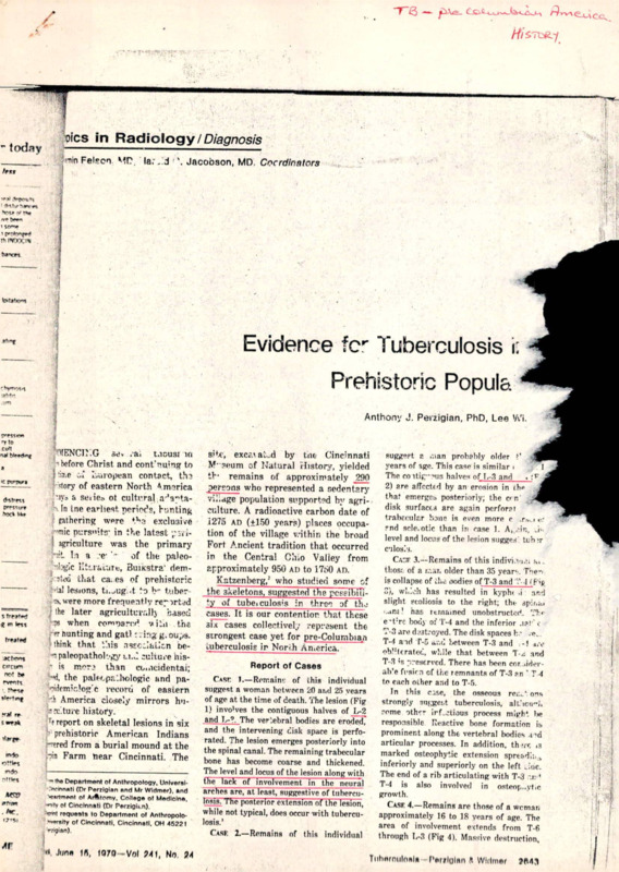
Evidence for Tuberculosis in a Prehistoric Population
Item
- Title
-
Evidence for Tuberculosis in a Prehistoric Population
- extracted text
-
flics in Radiology ZD/a^nos/s
”today
1
’•nin Felson MC. la.-, J 1 \ Jacobson, MD. Cocrdinators
fe<tt
T|
!
"J
■
> rtttir \rv«
rtosa v »•<•
•ve »v»-
1
th r*rrx_tv
bancM
kwtj'm-n
Evidence fcr Tuberculosis i:
ahnf
Prehistoric Popule.
' brmot-5
-ttm
Anthony J. Perzigian, PhD, Lee 'A'i.
3 '1ENCP.G adv ra. t.ious? m
T'tO
CuM
nal
p before Christ and conCnuing to
l*iiT.e
European contact, th3
dory of eastern North America
-ayo a seiiej. ot cultural a^a^taIn ine earliest, periods, hunting
fathering wore *>.-j exclusive
'mic ni:rs”’.ts- in the latest
agriculture was the primary
Ht In a -e__ the r
___
of
paleo’ilogic Ettracurfc, BuiKstra’ demnUa that ca. es of prehistoric
lesions, ti.uUg'.<t to b- tuber
were more frequently ref irted
the later agricultural!} based
when compared viui the
'«■ hunting and gat!: zing g. oups.
think that this nssc^fauon ben paleopathology li.J culture his- is more than cuacidentai;
•M, the pabcpF.Jwilogie and paoidemiclog'c reeoru of eastern
-Ji America closely mirrors hu•ev'.ture history.
rf report on skeletal lesions in six
prehistoric American Indians
rrered from a burial mound at the
loin Farm near Cincinnati. The
|_________
a
■c pinxi’i
<Ktr«»
bock kk*
_____
s
a m
aclxwn
ctrcucn
no! t»
rvontt
!b*^e
?<•* rr
5 weak
m<Jo
•om*t
>ndn
n!!lrt
■n the Department of Anthropolopy, Unlversl.•eeinnatl (Dr PerriQlan and Mr Widmer), and
•ortment of Arfttomy, Collepe of Medicine.
■'’tty of Cincinnati (Dr Perzipit.n).
•JTflt requests to Department of Anthropoloxersify of Cincinnati. Cincinnati. OH 45221
■rrijjian).
»Ptn
17’Sl
< Juno Ifl, 1070-Vol ?41, No, JM
1
site, excc.iau„d by tne Cincinnati
M-3oum of Natural History, yielded
th- remains of approximately 290
.persons who represented a sedentary
village population supported by agri
culture. A radioactive carbon date of
1275 ad (+150 years) places occupa
tion of the village within the broad
Fort .Ancient tradition that occurred
in the Central Ohio Valley from
approximately 950 ad to 1750 AD.
Katzenberg/ who studied some of
the skeletons, suggested the pcssibiuty of tubeiculosis in three of th-j
cases. It is our contention, that these
six cases collectively represent the
strongest case yet for pre-Columbian
tuberculosis in North America.
Report of Cases
Casc. 1.—Remain!: of this individual
suggest a woman between 20 and 25 years
of age at the time of death. The lesion (Fig
1) invo’ves the contiguous halves of L-2
and L-?. The vertebral bodies are eroded?
and the intervening disk space is perfo
rated. The lesion emerges posteriorly into
the spinal canal. The remaining trabecular
bone has become coarse and thickened.
The level and locus of the lesion along with
the lack of involvement in the neural
*rg^ea arc> at least, suggestive of tuhercu^sis. The posterior extension of the lesion,
while not typical, does occur with tubercu
losis.’
Case 2.—Remains of this individual
sugg?pf. a ,nan probably older t’ i
years of age. This case is similar i
The COltig-nua halves of 1,-3 and . IF
2) are affected by an erosion in rh^
4
that emerges posteriorly; the c»n’
disk surfaces are again performt
trabecular bane is even more c .r.^i ef
?nd scle.otic than in rase 1. A,/in, J.,
level and locus of the lesion sugges’ tuh>r
culos/j.
Cagf 3.—Remains of this indivi.’-uu .
those of a r.-.ar. older than 35 year1. Thei
is collapse of the oodies of T-3 and T 1 (Rig
3) , which has resulted in kyph< .i and
slight scoliosis to the right; the epina.
‘..ini' has itrnained unobstructed. Th*e't:re body o: T-4 and the inferior .i?! ‘c
T-3 are destroyed. The disk spaces h? <e..
T-4 and T-5 a.-.u between T-3 and -! a.v
ob’:krateG, while that between Tand
T-3 ?s -reserved. There has been cor ..iderab’e fi-sirn cf the remnants of T-3 an ' T-4
to each other and to T-5.
In this case, the osseous ref.v*. ons
strongly suggest tuberculosis, alficn"h
feme other irLctious process might be
responsible Reactive hone formation is
prominent along the vertebral bodies am
articular processes. In addition, th?rc ,3
marked osteophytic extension spread!:, j,
inferiorly and superiorly on the left. Joe.
The end of a rib articulating with T-3 -.i: '
T-4 is also involved in osteop,.yt.c
growth.
Case 4.—Remains are those of a woman
approximately 16 to 18 years of age. The
area of involvement extends from T-6
through 1,-3 (Fig 4). Massive destruction,
Tiitwoutofiln
PenHolan K Widmer
1
Ji
B
I1
g'
i
0 ;
I
i.-tB ■
tl
1
I
I1
I
■A
1
1
I
s
.
collapse, and severe kyphoscoliosis are
evident at T-8 toL-1. The bodies of T-6 and
T-7 are eroded posteriorly. Only T6-7, T78, and L2-3 disk spaces remain. The
collapsed bones also display extensive
fusion of the bodies and articular pro
cesses; several drainage channels are also
visible. A remnant of the right 11th rib is
fused to T-U. The remaining vertebrae,
L-4, L-5, and T-5 as well as some frag
ments of the cervical spine are free of
lesions. Both the level and degree of
destruction and collapse are strongly
suggestive of tuberculosis.
Case 5.—Remains are those of a woman
between 16 and 18 years of age. The bodies
of C-4 through T-2 show destruction and
collapse (Fig 5); C-2, C-3, and T-3, though
involved in the fusion, have intact bodies.
The collapsed vertebrae exhibit a double
scoliosis oriented to the right from T-2 to
about C-7 and then to the left to C-2.
Kyphosis is severe; however, the spinal
canal is unobstructed. All disk spaces from
C-2 to T-2 are obliterated. There is
massive fusion but with no osteophytic
spurring. Posterior fusion of the articular
and spinous processes is more marked on
the left side than on the right. While a
locus of tuberculosis in the cervical and
upper thoracic spine is not common, it is
not unknown. This case, with its destruc
tion of the bodies, consequent collapse, and
subsequent ankylosis, clearly rusemblcs
case 3 and particularly case 4.
Case 6.—Remains suggest a woman. A
lesion is centered in the remnants of the
left acetabulum (Fig 6). Much of the corti
cal bone is destroyed. A drainage canal
penetrates the upper lateral side of the
acetabulum and emerges on the medial
surface of the ilium (arrows). Only the
shaft of the left femur remains. The proxi
mal portion displays medial periosteal
reaction that extends to the linea aspera;
presumably, the missing metaphysial and
articular portions were even more severely
compromised. The right acetabulum and
femur were unaffected. This unilateral
lesion of the acetabulum and proximal
femur is highly suggestive of tuberculo
sis.
Comment
■
1
i
? •1
$
Bll
irB If
Despite the uncertainties in paleopathologic diagnosis, these six cases
present strong testimony favoring the
presence of tuberculosis in preColumbian North America. In a
sedentary village population like Tur
pin, where tuberculosis is endemic, we
would expect osseous lesions to occur
preponderantly in the thoracolumbar
vertebrae and less commonly in the
cervical spine or hip. The pattern
reported here is compatible with
these expectations. Cases 1 through 5,
2644
r'*-'
flFkj 3.—
Fig 1.—Case 1. Superoinferior roentgeno
gram of L-2 (top) and L-3 (bottom)..
Fig 2.—Case 2. Superoinlenoc roarAjai'.
gram of L-3 (top) and L-4 (botioro)
moreover, follow the classic pattern
of tuberculosis spondylitis; each individual suffered lytic destruction of
either the center or the anterior
portion of the vertebral bodies or
bothT In addition, the number of
*ankylosed vertebrae and the neural
arch involvement are not inconsistent
with this diagnosis? In the healed
stage, as few as two or as many as
eight or more vertebral bodies may
form one tuberculous block vertebra.
Kyphosis is also not uncharacteris
gested infection at a much
age, followed by remission,
and ankylosis.
The severe vertebral dcstr.x'uu
jnight also suggestL bacteriaLfiCai
cotic infection. Actinomyco»i» om
probably be ruled out. The cenx>
facial region is most cuma*.^
involved.0 Actinomycosis rartl>
to collapse of vertebral bodies, I-wa
or angulation of the spine.*4
Spinal blastomycosis on the cOur
hand, does show a predeliction (<* u*
thoracolumbar region’ and is
in the Ohio River Valley. Furu<r
more, Buikstra1 has suggested tu.
exposure to a soil-borne fungus
Blastomyces dermatilidis prubabtj creased with the adoption of agr-iu*ture by aboriginals. This would u«a
conveniently explain the much Lgsxr
incidence of vertebral lesions in
farming groups when compared a&k
earlier hunters and gatherers.
We believe, however, that Uhtra
losis is a better explanation for Os
tic.
We might anticipate that both
subadults and adults would display
such lesions. Unfortunately, sub
adults were underrepresented, ac
counting for only 42 of the 290 indi
viduals we examined. Perhaps not
coincidentally, no lesions resembling
tuberculosis were recorded in this age
group. However, three of the five
individuals with spinal involvement
were women approximately 20 years
old or younger whose lesions sug-
JAMA, June 15, 1979—Vol 241, No. 24
Tuberculosis—Perzigian & w^ja.
Ftg 5.—
(nght) o
JAMA,
3 i
Vi :-4,
w
3^
,T-3
■
' <'•
'V’
'?■
41
h
../hh
j *
fe
-
•./
-
$
-
' •
■i
V
I
•■V/
■.
h■
h'
t .' ''’'I •’l- fi‘ j ’ '• ” '
-Case 3. Lateral roentgenogram of T-3, T-4, and T-5.
r
$
>r roentg- •
tom)
Fig 4.—Case 4. Lateral roentgenogram of T-8 through L-3.
ch ynur .• •
m. bonV • r ‘,-Case 5. Posteroanterior (left) and lateral roentgenogram
“I of C-2 through T-3.
rh-»r- • •
rial or
yewtiw
ywrwjR
r‘
■
ci
I >1I
he rer". •'
cornr-' ■
rarely l» 1
•Jies, f'liv
T J V>r
• ■
L.
i.-i
tinn fo* •
<
’v
1'"^.
’ 4 %•*1 < '•
>
1
j
^4of >f’
-
,. ..
Mw
wntjlc
• • _* kk
i
inch *• r * ■’•
•
•■
‘
h
■
i
~ • -• ;
jpfl’. 1
-
■
funtr’s
•
■
■ fiU
i is r- ’• y. Furl*”
-’C'te,!
Fig 6.—Case 6. Roentgenogram of remnants (arrows) of left
acetabulum.
■.
<
■
'-r
’W;
k
'em
at tv‘^-
5<»n 1 • ■ ‘ •
nn * V»
__________
ItlHH ff>,
10/0
Vol SMI, No
34
TiihtiroiiloHlH
Parilolan A Wiiiinm
?H4fi
* Jir
n'l
findings than blastomycosis. The an
kylosis and extreme kyphosis of our
cases are not typical of advanced
stages of disseminated blastomycosis.
Untreated
blastomycosis
rarely
reuchuM u Hubclinicul stugu with spon
taneous healing.*"' Blastomycosis, a
rare disease, is not known to be trans
mitted from person to person"; rath
er, spores are first inhaled and only
later spread through lymphatic and
hematogenous dissemination. At the
Turpin site, the similarity of the
lesions and the number of individuals
affected along with the apparent
'temporal and spatial proximity of the
"burials all suggest the presence of an
endemic highly communicable pathotgen.
Juvenile
rheumatoid
arthritis
(Still’s disease) is suggested by
Katzenberg* as the diagnosis in case
5. It is typically a diffuse systemic
disease with considerable peripheral
joint involvement12 and severe growth
disturbance of the long bones."
Growth arrest lines are common."
The spine is most often fused at C2-3
and less often at C3-4 or lower.'*'1*
Case 5 exhibits none of the classic
characteristics of juvenile rheuma
toid arthritis. Peripheral joints ex
hibited no arthritic pathology; long
bones were free of growth arrest
lines. Furthermore, the involvement
of C-5 through T-3 is hardly charac
teristic of juvenile rheumatoid arthri
tis. We find more compelling support
for tuberculosis, despite the cervical
location.
Within the last 8,000 years, the
virtual abandonment of hunting and
gathering as a previously universal
life-style had a profound global effect
on prehistoric settlement patterns,
social relations, technology, and diet.
In eastern North America, for exam
ple, we can see important demograph
ic changes as farming villages arose
and increased in number and size.”
Local population densities reached
unprecedented levels; dependence on
j:orn agriculture grew accordingly.
The disease profiles of prehistoric
1. Buikstra JE: Differential diagnosis: An
epidemiologic model. Yearbook Phys Anthropol
20:316-328, 1976.
2. Rauenberg MA: An investigation of spinal
disease in a midwest aboriginal population.
Yearbook Phys Anthropol 20:349-355, 1976.
3. Schmorl G, Junghanns H: Human Spine in
Health and Disease, ed 2. New York, Grune &
Stratton Publishers, 1971, p 314.
4. Rosen RS, Jacobson J: Fungus disease of
bone. •Semin iCoenlyvnul 1:370-391, 1966.
5. Goldman AB, Freiberger Rii: Infectious
and neuropathic localized diseases of the spinal
column. Semin Roentgenol 14:19-32, 1979.
6. Flynn MW, Felson B: The roentgen mani
festations of thoracic actinomycosis. Am J
Roentgenol 110:707-716, 1970.
7. Jaffe Hk Metabolic, Degenerative, and
Injtammatory Diseases of Bones and Joints.
Philadelphia, Lea & Febiger Publishers, 1972, p
1066.
8. Tabb JL, Tucker JT: Actinomycosis of the
spine. Am J Roentgenol 29:628-634, 1933.
9. Curtis AC, Florante CB: North American
blastomycosis. J Chronic Din 5:404-429, 1957.
10. Martin DS, Smith DT: Blastomycosis: 11. A
report of 13 new cases. Am Rev Tuberculosis
39:488-515, 1939.
11. Schwarz J, Baum GL Blastomycosis. Am J
Clin Pathol 21:999-1029, 1951.
12. Brewer EJ: Juvenile Rheumatoid Arthri
tis. Philadelphia, WB Saunders Co, 1970.
13. Martel W, Holt JF, Cassidy JT. Roentgeno
logic manifestations of juvenile rheumatoid
arthritis. Am J Roentgenol 88:400-423, 1962.
14. Ansell BM, Bywaters EGL Growth in
Still's disease. Ann Rheum Dis 15:295-319,1956.
15. Ansell BM: The cervical spine in juvenile
•;W
populations changed comc^auu.- ua
ly. Indeed, tuberculoti»-Ui4 mmu.a*
reported here and
actually confirmed in a
bian Peruvian mummy'w .
confined to sedentary
. .. u.
based communities. Ou Uw ium*
hand, earlier hunting gruup* •<r«. a*
doubt, much less exposed u <n*l
infections like tul*4ulusu -U in.
bos19 emphasizes, n«<i tuuL ua
technological changes
«.
ther immediate or delayed pfiy n
cal disturbances and nuy ar: w
direct or indirect causes of tUuM
This paleopathologic study, u-n,
reaffirms this view and ren^kj m
that sociocultural change is yr-«au>
nent * in determining epidz-^^u
patterns.
Edward H. Miller, MO, ar.J Uxw
port, MD, of the University of Ciw.ixu.
of Medicine, and Paul Jolly, MD.uf n* ;uu«..u^
County Coroner’s Otfice, aided n u*
lion of the patholo|£y sludie* TU
u—
rial was made available by u< Caa immm
Museum of Natural History
'* <□•»*
assisted in the preparation of lh<
References
■ Ur
4
.f
New Appointments
Harold G. Jacobson, MD, new editor for the TOPICS
in radiology section of JAMA, has appointed two
senior coordinators for Diagnostic Radiology, Jack
Edeiken, MD, and E. Robert Heitzman, Jr, MD, both
outstanding men in the field.
Dr Edeiken, professor of radiology at Jeffer.on
Medical College and chairman of the Department of
Radiology, Thomas Jefferson University Hospital,
Philadelphia, has concentrated on orthopedic radiol
ogy. Author of Roentgen Diagnosis and Diseases of
Bone (Baltimore, Williams & Wilkins Co, 1967; ed 2,
1973; Spanish ed, 1977) and Roentgen Atlas of the
Hand and Wrist in Systemic Disease (Baltimore,
Williams & Wilkins Co, 1973), Dr Edeiken has also
published more than 60 journal articles and syllabi
and is co-editor-in-chief of Skeletal Radiology. He
3
rheumatoid arthritis, in Carter Mt >«u.
logical Aspects of Rheumutuul
Pn
ceedings of an Internutumai SymyuesuM. ->~.
national Congress Scries Nu al
!<•<.
Excerpta Medica FoundaUun. IM, ? SK
16. Ziff M, Conlreras V, M<E.«n C v— •
tis in post-pubertal patients wita
arthritis of juvenile onset.
kA»u» Im
45, 1956.
17. Ford RI: Northeastern *rri«.«.<_ l<u
and future directions.
Juv
mm
3:3ij5-4l3, 1974.
K18. Allison MJ, Mendoza D. Pczua I .»■
mentation of a case of lubcrcuio^* a »>»
Columbian America. Am Rev
Im sit
991, 1973.
rt9yDubos R: Man, Mcdicnu, aaJ
ment London, Pall Mall Pre**, p Tt
serves as consultant for radiology journals and u a
member of many committees in radiological socie
ties and associations.
Dr Heitzman is director of the Diagnostic Divi
sion, Department of Radiology, at the State Univer
sity of New York Upstate Medical Center, Syracuse.
He is an outstanding authority on radiology of chest
diseases. He has served on several committees <J
radiological societies and associations and has he4
many offices, including the presidency of Th*
Fleischner Society, 1978-1979. Dr Heitzman is aa
editorial board member of Investigative Radiol^t
and American Journal of Roentgenology and Lu
published more than 50 articles and many syllat*
and exhibits. He is co-editor of An Atlun of CruuSectional Anatomy (New York, Harper 4 Ru»
Publishers Inc, 1979).
■i!
ir'ft
-
f
2646
JAMA. June 15. 1979—Vol 241. No. 24
Tuberculosis—PerziQan A
Rl
IB
J-w
- Media
 RF-TB-1.27.pdf
Position: 1294 (16 views)