History and Importance of Scrofula
Item
- Title
- History and Importance of Scrofula
- extracted text
-
MMtt • l
THE LANCET
Department of medical history
History and importance of scrofula
Stefan Grzybowski, Edward A Allen
!
The clinical entity called scrofula has for many centuries,
by its highly visible characteristics, provided a marker for
tuberculosis.
In his book A Handbook of Geographical and Historical
Pathology, August Hirsch1 provided a picture of scrofula as
it was understood until the discovery in 1882 of the
tubercle bacillus by Robert Koch. The term scrofula, he
stated, “denotes an inflammatory kind of tumour, more
particularly in the neck”. The word is a diminutive of the
Latin word scrofa, a breeding sow supposedly prone to
the disease, which was recorded by Aristotle.2 The word
corresponds etymologically to the Greek for pig, but
Hirsch questioned whether it should not be taken in the
figurative sense meaning a stone, reflecting the scirrhous
hardness of the lymph glands when inflamed, as described
by Galen. The word scrofula is first encountered in
medical writings of the school of Salerno, Italy, in the
early 16th century, but it first became a “technical term”,
as Hirsch puts it, in the Hippocratic period in Greece
when the condition was especially common in children
and its protracted course and “cold and mucous nature”
were identified. The lack of knowledge about its microbial
cause hampered final confirmation of its unity with other
forms of tuberculosis until the end of the 19th century.
Despite Koch’s demonstration of tubercle bacilli in
scrofulous lymph nodes, some writers remained
unconvinced, either on epidemiological grounds or on the
basis of human inoculation experiments, that scrofula was
transmissible; this led them to deny a link with
tuberculosis and thus to reject the unity of scrofula and
pulmonary tuberculosis. Even William Osler, convinced
as he was by the work of Koch, wrote “It is not yet
definitely settled whether the virus which produced the
chronic adenitis or scrofula differs from that which
produced tuberculosis in other parts”.’
In this account, we consider three aspects of the history
of scrofula—the King’s Evil, Marfan’s law, and the place
of this type of tuberculosis in the epidemiology of this
disease.
Daniel5 suggested that “royal claims of a cure might have
gained widespread acceptance” because “tuberculosis
adenitis usually represents primary infection with often a
benign course and that primary infection often confers
immunity”.
The formal practice of the ceremonial rite can be traced
back to the reigns of St Louis (Louis IX 1226-70) in
France and Edward III (1327-77) in England. Edward III
was the first English king to order a public display of this
rite; he used a medallion called a touch-piece, which was
given to sufferers as a sort of talisman? Initially, the
ceremony consisted of the king washing the diseased flesh
with water, but Henry VII discontinued this practice.
Instead, the ceremony consisted of the king touching the
afflicted subject while the court chaplain recited prayers
and presented the touch-piece, which was usually
suspended by a silk ribbon around the neck. Edward I
(1272-1307) touched 533 of his subjects in 1 month;
Philip VI of Valois (1328-50) touched 1500 at a single
ceremony; Charles II (1660-82) according to the registry,
touched 92 102 people during his 22-year reign, at times
600 in one ceremony. Louis XVI, soon to meet the
guillotine, was anointed at his coronation with the Holy
Oil of Clovis on June 11, 1775, and “three days later in
the summer, he ritually touched 2400 stinking sufferers
from scrofula”.7 William III (1689-1702) allowed the
ceremony to lapse after a single performance with the
remark to the ailing people, “God give you better health
and more sense”. He refused to touch the patients but
.-’I
1
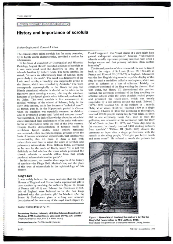
King’s Evil
It was widely believed for many centuries that the Royal
Houses of England and France had a supernatural gift to
cure scrofula by touching the sufferers (figure 1). Clovis
of France (481-511) and Edward the Confessor (104266) of England were believed to be the first kings
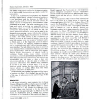
endowed with this particular gift.4 In Macbeth (Act IV,
Scene 3) Shakespeare gives a striking and accurate
description of the ceremony of the royal touch (figure 2).
'I
I
Lancet 1995; 346: 1472-74
1
<
Respiratory Division, University of British Columbia Department of
Medicine, 2775 Heather Street, Vancouver, BC V5Z 3J5, Canada
(Prof S Grzybowski frcp, Prof E A Allen frcpc)
Correspondence to: Prof Stefan Grzybowski
1472
~ } .J....
.
Figure 1: Queen Mary I touching the neck of a boy for the
King's Evil (watercolour by M S Lapthom, 1911)
Reproduced with permission of Wellcome Institute Library, London,
Vol 346 • December 2, 1995
THE LANCET
Enter a doctor
Malcolm Well; more anon—Comes the king forth, I pray
- you?
Doctor
Ay, sir; there are a crew of wretched souls
That stay his cure; their malady convinces .
. . The great assay of art; but at his touch—
Such sanctity hath heaven given his hand—
They presently amend.
Malcolm I thank you, doctor
[Exit Doctor
Macduff What’s the disease he means?
Malcolm ‘Tis called the evil: T - ‘
:
A most miraculous work in this good king;
Which often, since my here-remain in England,
I have seen him do. How he solicits heaven,
Himself best knows: but strangely-visited people,
All swoln, and ulcerous, pitiful to the eye, . ..
The mere despair of surgery, he cures,
Hanging a golden stamp about their necks,
Put on with holy prayers; and ‘tis spoken,
To the succeeding royalty he leaves
The healing benediction. With this strange virtue,
He hath a heavenly gift of prophecy,
And sundry blessings hang about his throne,
That speak him full of grace.
Figure 2: Passage from Macbeth (Act IV, Scene 3) describing
the ceremony to cure the King's Evil
referred them to the exiled James II in France. The
practice was continued by the exiled Stuarts and was
frequently done in Italy by James Stuart, “the old
pretender”, and by his two sons, Charles and Henry
(Cardinal of York). Samuel Johnson was one of the last
sufferers of scrofula to be touched. As a child, he was
taken to Queen Anne (1702-14). His step-daughter
recalls that he wore his touch-piece proudly into
adulthood along with the scars on his neck.8
Marfan remembered
In 1886, Bernard Jean Antonin Marfan (1858-1942)
stated that individuals with scars from lupus and scrofula
rarely develop phthisis (pulmonary tuberculosis)? This
clinical observation came to be known as Marfan’s law.
Support for Marfan’s observation came from Von
Pirquet10 and Fowler." Osler’ doubted the validity of his
law. The question of whether Marfan’s law was true or
false remained controversial for many decades until the
work of Francis in Britain,12 Magnus in Denmark," and
Sjogren and Sutherland in Sweden14 revealed that
pulmonary tuberculosis was less common in communities
with a high prevalence of tuberculosis in cattle than in
those with less bovine infection.
The bovine tubercle bacillus Mycobacterium bovis has
been a common cause of cervical adenitis in human
beings in countries with a high prevalence of tuberculosis
in cattle. In England, for instance, as late as the 1940s
and 1950s, M bovis was responsible for 57-5% of cases of
cervical adenitis but for only 1-2% of cases of pulmonary
tuberculosis.” Although the cause of the adenitis observed
by Marfan is not known, it is reasonable to suppose that
most of the cases were caused by the bovine strain in the
light of subsequent epidemiological studies.
It is now clear that cervical adenitis may arise in two
distinct ways. Cervical lymph nodes may be a part of the
primary complex when the primary focus is located in the
tonsils and pharynx, as happens in infection with bovine
tubercle bacilli spread by contaminated milk. The
alimentary route of infection may also lead to the
glandular component of the primary complex in the
Vol 346 • December 2, 1995
mesenteric lymph nodes. Although human disease as a
result of bovine infection is now rare, it may be serious
and even fatal.
Cervical adenitis due to
tuberculosis is a feature of the
spread of infection from the lungs, usually (or perhaps
almost always) through the haematogenous route, though
lymphatic pathways have been proposed.'6 This type of
cervical adenitis is serious because it indicates that there
are tuberculous foci in the lungs and that there may be
other haematogenous foci in the body.
Cervical adenitis may also be due to non-tuberculous
(environmental) mycobacteria, in most cases M aviumintracellulare or M scrofulaceum.'' This illness occurs in
children in areas where bovine infection has been
eradicated and human tuberculosis has become rare and
where appropriate environmental mycobacteria are
prevalent."1 In British Columbia, cervical adenitis due to
atypical mycobacterial infections has become the most
common form of mycobacterial adenitis in childhood."
Epidemiological importance of cervical
adenitis
Tuberculous lymphadenitis accounts for some 5% of total
active cases of tuberculosis reported in Canada.20 It is
unevenly distributed in the population, being especially
common in immigrants from Asia, in whom this form of
disease accounts for about one-quarter of all active cases.
In Canada, cervical adenitis is also more than twice as
common among Indians and the Inuit than in the white
Canadian-born population.2' In all these groups, the
disease is mainly caused by Af tuberculosis with M bovis
accounting only for about 1% of cases.20
In a study of tuberculosis in British Columbia among
immigrants from five Asian countries between 1982 and
1985,22 tuberculous adenitis accounted for 24% of total
active cases, varying from 10% for those born in Japan to
44~% for those born in the Philippines. The high
frequency of cervical adenitis in immigrants from Asia has
been found elsewhere. In the UK, a survey in 1971 by the
British Tuberculosis and Thoracic Association showed
that lymphadenitis accounted for 25-3% of all
tuberculosis notifications in patients born in India and
Pakistan.2’ The Scottish National Survey of tuberculosis
notifications in 19932' confirmed a relatively high rate of
non-pulmonary disease in immigrants from the Indian
subcontinent (47%), with lymphatic disease making by far
the largest contribution (67%).
Discussion
Tuberculous cervical adenitis has afflicted mankind
probably for thousands of years. That royalty was believed
to possess supernatural powers over this disease points to
its frequency over the past 1500 years. However, a more
serious study of all the historical documents relating to
the royal touch might provide us with meaningful
epidemiological data on this type of tuberculosis. By
contrast, the occurrence of pulmonary tuberculosis was
not often mentioned before the middle of the 15th
century. Descriptions of phthisis by Hippocrates and by
others from antiquity leave open the possibility that
aetiologically different, yet clinically similar, wasting
diseases were included under this title. Such
considerations accord with Bates’ and Stead’s suggestion
that the human tubercle bacillus has evolved only during
the current millennium.25
1473
THE LANCET
Marfan’s law was scarcely referred to by Marfan’s
contemporaries and has been largely forgotten, although
the name of this astute French paediatrician is
remembered by many medical students for the syndrome
that he described. In many countries it took decades to
show that cervical adenitis is often of bovine origin, and a
similar period to show that infection with M bovis,
acquired usually through contaminated milk, is protective
against respiratory infection with the human tubercle
bacillus. Perhaps the rapid and successful attempts to
control and virtually eradicate bovine infection when the
onslaught of infection with M tuberculosis had peaked was
a mixed blessing: human infection with the bovine strain
was an unintended and dangerous, yet potent,
vaccination. In retrospect, it is not surprising that
Marfan’s law remained controversial for so long.
Clinicians whose patients with cervical adenitis were
predominantly infected by bovine bacilli were convinced
of its truth, whereas those who dealt mainly with adenitis
caused by the human bacillus were, like Osler,’ rather
doubtful.
Non-tuberculous mycobacteria seem to have replaced
the bovine bacillus both as a cause of cervical adenitis”
and as a natural vaccinating agent.26 Infections with these
organisms act as a rather weak vaccine with few
complications in individuals with a normal immune
response.
The reason for the high frequency of adenitis in
immigrants from Asia is not completely clear; it is
probably, at least partly, the expression of the tuberculosis
epidemic in these groups at an earlier stage than that seen
in Europeans. Lin,27 in his thoughtful analysis of the
tuberculosis problem in East Asia and the South Pacific
area, suggested that the high frequency of extrapulmonary
tuberculosis and of tuberculosis in children, and the
almost equal incidence of tuberculosis in males and
females are all suggestive of an earlier stage of the
tuberculosis epidemic. Cummins, in his monograph
Primitive Tuberculosis,23 provides plenty of evidence of the
high frequency of lymphadenitis in the early stages of the
epidemic.
AIDS will open a new chapter in the story of scrofula.
Not only can HIV itself cause lymphadenopathy, but also
the loss of immunity allows for both the recrudescence of
latent mycobacterial foci in the glands and the acquisition
of new infections.
We thank Ms Cecile Russell for excellent secretarial assistance.
References
1 Hirsch A. A handbook of geographical and historical pathology. Vol II
(translated from 2nd German ed by Creighton C). London: The New
Sydenham Society, 1885: 604-41.
2 Keers RY. Pulmonary tuberculosis: a journey down the centuries.
London: Bailliere Tindall, 1978: 6-19.
1474
3 Osler W. The principles and practice of medicine. New York:
D Appleton and Company, 1892: 184-256.
4 Encyclopaedia Britannica, 11th ed. Cambridge: Cambridge University
Press, 1910.
5 Daniel TM, Bates JH, Downes KA. History of tuberculosis.
In: Bloom BR, ed. Tuberculosis: pathogenesis, protection, and control.
Washington DC: American Society for Microbiology Press, 1994:
13-24.
6 Spalding M, Welch P. Nurturing yesterday’s child: a portrayal of the
Drake collection of paediatric history. Philadelphia: B C Decker, 1991:
175-89.
7 The Oxford history of the French revolution. Oxford: Oxford
University Press, 1989: 1.
8 Hardy JP. Samuel Johnson: a critical study. London, Routledge and
Kegan Paul, 1979: 27-34.
9 Marfan A. De I’immunite conferee par las guerison d’une tuberculose
locale pour la phthisic pulmonairc. Arch Gen de Med 1886; 57: 575.
10 von Pirquet C. Tuberculosis in childhood. In: Klebs AC, ed.
Tuberculosis: a treatise by American authors on its etiology, pathology,
frequency, semeiology, diagnosis, prognosis, prevention, and treatment.
New York: D Appleton and Company, 1909: 147.
11 Fowler KJ. Pulmonary tuberculosis. London: MacMillan, 1921: 3-44.
12 Francis J. Control of infection with the bovine tubercle bacillus. Lancet
1950; i: 34-39.
13 Magnus K. Epidemiological basis of tuberculosis eradication: risk of
pulmonary tuberculosis after human and bovine infection. Bull World
Health Organ 1966; 35: 483-508.
14 Sjogren 1, Sutherland I. Studies of tuberculosis in men in relation to
infection in cattle. Tubercle 1974; 56: 1 13-27.
15 Wilson GS, Blacklock JNS, Riley RW. Non-pulmonary tuberculosis of
bovine origin in Great Britain and Northern Ireland. London: National
. Association for Prevention of Tuberculosis, 1952.
\J6 Miller FJW, Cashman JM. Origin of peripheral tuberculous
lymphadenitis in childhood. Lancet 1958; i: 286-89.
17 Wolinsky E. Nontuberculous mycobacteria and associated diseases.
Am Rev Respir Dis 1979; 119: 107-59.
18 Allen EA. Tuberculosis and other mycobacterial infections of the lung.
In: Thurbeck WM, Churg AM, eds. Pathology of the lung, 2nd ed.
New York: Thieme Medical Publishers, 1995: 253-54.
19 Robakiewicz M, Grzybowski S. Epidemiological aspects of
nontuberculous mycobacterial disease and of tuberculosis in British
Columbia. Am Rev Respir Dis 1974; 109: 613-20.
20 Enarson DA, Ashley MJ, Grzybowski S, Ostapkowicz E, Dorken E.
Non-respiratory tuberculosis in Canada. Am J Epidemiol 1980; 112/3:
341-51.
21 Brancker A, Enarson DA, Grzybowski S, Hershfield ES, Jeanes CWL.
A statistical chronicle of tuberculosis in Canada. Health Rep 1992; 4.
Catalogue 82-003:20.
22 Wang JS, Allen EA, Chao CW, Enarson D, Grzybowski S. Tuberculosis
in British Columbia among immigrants from five Asian countries 19801985. Tubercle 1989; 70: 179-86.
23 British Thoracic and Tuberculosis Association. A tuberculosis survey in
England and Wales, 1971. The influence of immigration and country
of birth upon notifications. Tubercle 1973; 54: 249-60.
24 Leitch AG, Rubilar M, Forbes GI, et al. Scottish national survey of
tuberculosis notification in 1993 with special reference to the
prevalence of HIV seropositivity. Thorax 9195; 50: 442P.
25 Bates JH, Stead WW. The history of tuberculosis as a global epidemic.
Med Clin North Am 1993; 77: 1205-70.
26 Palmer LE, Long MW. Effects of infection with atypical mycobacteria
on BCG vaccination and tuberculosis. Am Rev Respir Dis 1966; 94:
553-68.
27 Lin HT. The tuberculosis problem and its control in East Asia and the
South Pacific area. Bull IUAT 1986; 61: 28-39.
28 Cummins SL. Primitive tuberculosis. London: John Bale Medical
Publications, 1939: 47-49.
Vol 346 • December 2, 1995
- Media
 RF-TB-1.15.pdf
Position: 170 (127 views)