Tuberculosis: Pathogenesis, Protection, and Control
Item
- Title
- Tuberculosis: Pathogenesis, Protection, and Control
- extracted text
-
Tuberculosis: Pathogenesis, Protection, and Control
Edited by Barry R. Bloom
© 1994 American Society for Microbiology, Washington. DC 20005
Chapter 32
Strategies for New Drug Development
Douglas 13. Young
The emergence of strains of Mycobacte
rium tuberculosis resistant to existing drugs
has focused attention on the urgent need for
development of new antimycobacterial
agents. Such agents have not been per
ceived as a high priority by pharmaceutical
companies over the last 30 years, and a
coordinated effort to screen general antimi
crobial compounds developed during this
time for activity against M. tuberculosis
may well prove worthwhile. The recent
development of genetic tools for monitoring
the viability of M. tuberculosis provides a
rapid approach for this type of screening
(Jacobs et al., 1993). From a broader per
spective, molecular genetic tools for study
and manipulation of mycobacteria provide
access to a vast amount of new information
about the biochemistry and metabolism of
M. tuberculosis, and exploitation of this
information has important potential in the
rational development of a new generation
of antimycobacterial agents and perhaps in
the design of improved strategies for use of
existing drugs. This chapter focuses on the
prospects for using a fundamental molecu
lar approach to identification of novel lead
compounds for new drug development.
Further important steps in drug develop
Douglas B. Young • Department of Medical Micro
biology, St. Mary’s Hospital Medical School, Norfolk
Place, London W2 IPG, United Kingdom.
559
ment, such as toxicity testing, optimization
of pharmacokinetics, etc., are not ad
dressed in this review.
In selecting targets for antimicrobial
agents, it is clearly advantageous to avoid
bacterial enzymes with closely related
counterparts in mammalian cells. In addi
tion, to avoid disruption of normal micro
bial flora during the prolonged course of
tuberculosis therapy and to limit possible
transfer of resistance factors from other
bacteria! genera, it is preferable that new
drug targets be specific for mycobacteria.
Drugs must act on a target that is essen
tial for bacterial survival, and ideally,
they should be effective against bacteria
throughout their growth cycle both inside
and outside mammalian cells during in
fection. In this section, we first review
existing and potential drug targets in M.
tuberculosis. We then discuss distinctive
features of mycobacteria relevant to drug
design, and finally, we consider experimen
tal approaches applicable to rational drug
discovery programs.
DRUG TARGETS IN M. TUBERCULOSIS
Most antibacterial agents inhibit biosyn
thetic pathways involved in the production
of macromolecules (proteins, nucleic acids,
or cell wall polymers). Several of the broad
spectrum antibacterial agents are effective
560
Young
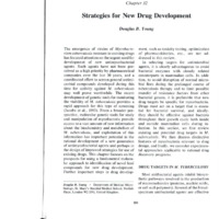
ONA gyrasc
(quinolones)
HNA polymerase
(ritampicin)
to be effective against tuberculosis suggest
that the ribosome may not be a particularly
attractive target for new antituberculous
drug design.
Nucleic Acids
■•iranydrololala
folate
metabolism
(PAS.
dapsone ■>)
rlbosome
(streptomycin)
cycloserine: peptidoglycan synthesis
isoniazid: mycollc acid synthesis?
cell wall biosynthesis ethambutol: arablnogalactan/
arabfnomannan synthesis?
ethionamide : mycollc acid synthesis?
Figure 1. Sites of action of antimycobacterial agents.
PABA, p-aminobenzoic acid; DHFR, dihydrofolate
reductase; PAS, p-aminosalicylic acid.
against mycobacteria (Fig 1) and the sites
•
g lh dnd the S,tes
of action of these existing drugs clearly
represent potential targets for new drug
development.
Protein Synthesis
Streptomycin, the first antibiotic avail
able for widespread use in treatment of
tuberculosis, is a member of the aminogly
coside family that disrupts bacterial protein
synthesis. As in other bacteria, streptomy
cin resistance in M. tuberculosis is conferred by mutations that alter the ribosomal
protein S12 or the ribosomal 16S RNA
molecule (Finken et al., 1993). Kanamycin,
a related aminoglycoside with a similar
mode of action, and its semisynthetic deriv
ative amikacin are also used in tuberculosis
therapy. While protein synthesis is clearly
an important drug target in M. tuberculosis,
other families of protein synthesis inhibi
tors (tetracycline, chloramphenicol, and
the macrolides [e.g., erythromycin)) have
no clinical use against tuberculosis. The
intensive effort that has gone into develop
ment of protein synthesis inhibitors and the
rather limited spectra of those agents found
LL
Sulfonamides, which were the first clini
cally effective antibacterial agents, are
structural analogs of p-aminobenzoic acid
that inhibit biosynthesis of tetrahydrofolic
acid, thus blocking production of the purine
and pyrimidine bases required for nucleic
acid synthesis. The antituberculous drug
p-aminosalicylic acid (Fig. 2) was initially
designed as a competitive inhibitor of sali
cylic acid and may act on the tetrahydrofo
late pathway as well as on the salicylatedependent biosynthesis of mycobactins
-hat
reqUired for iron transport. An
'mportant strategy for enhancing the activ
ity of sulfonamides against some bacteria
has been their use in combination with
trimethoprim, a drug that inhibits a subse
quent step in the tetrahydrofolate pathway
catalyzed by the enzyme dihydrofolate re
ductase. Although trimethoprim is not ac
tive against mycobacteria, a considerable
amount of structural information is avail
able concerning bacterial and mammalian
dihydrofolate reductases, and a detailed
study
of M.’ituberculosis vuz-ymes
enzymes from
V/'r
uom ithe
letrahydroto,ate pathway may provide: a
H\/°
CjH5—CH —NH—CHj—CHj—NH—CH—CjHs
CHjOH
CH,OH
NH,
ethambutol
p-amlnosallcylic acid
(PAS)
^NH-NH,
V"Hi
„
'’oNH)111
-CH
CHa3—
CH3
ethionamide
ethionamide
pyrazinamide
Figure 2. Structures of antimycobacterial
agents.
chapter 32
rculosis suggest
be a particularly
antituberculous
S
e the first clinial agents, are
nobenzoic acid
tetrahydrofolic
on of the purine
red for nucleic
berculous drug
2) was initially
nhibitor of salie tetrahydrofothe salicylatef mycobactins
transport. An
icing the activsome bacteria
nbination with
ihibits a subsefolate pathway
hydrofolate reorim is not aca considerable
lation is availid mammalian
nd a detailed
ymes from the
lay provide a
-CHj— NH—CH— C,H5
CH,OH
butol
pyrazlnamlde
acterial agents.
basis for rational design of novel synergistic
drug combinations.
The fluoroquinolones (ofloxacin, cipro
floxacin, sparfloxacin) are broad-spectrum
antibacterial agents that disrupt the bacte
rial chromosome by inhibiting the super
coiling activity of DNA gyrase. The fluoro
quinolones are increasingly important in
treatment of mycobacterial diseases, and
the genes encoding both subunits of the
DNA gyrase enzyme of M. tuberculosis
have been cloned (Takiff, personnal com
munication). Pharmaceutical companies
have invested considerable effort in devel
opment of gyrase inhibitors, and it may be
of interest to screen for evidence of speci
ficity for the mycobacterial enzyme among
such compounds.
Rifampin is a key drug in mycobacterial
therapy that has a broad antibacterial spec
trum and a well-defined target. In this case,
transcription is inhibited by an interaction
with the p subunit of the bacterial RNA
polymerase molecule. In mycobacteria, as
in other bacteria, resistance is conferred by
point mutations in the rpoB gene (Telenti et
al., 1993).
Cell Wall Biosynthesis
Broad-spectrum antibacterial agents
Biosynthesis of cell wall peptidoglycan
provides targets for a large number of anti
bacterial agents. Cycloserine inhibits incor
poration of o-alanine into the peptidogly
can precursor, while the glycopeptide drugs
vancomycin and teicoplanin inhibit assem
bly of the precursors by binding to the
terminal D-Ala-D-Ala residues. Members
of the extensive family of p-lactams (peni
cillins and cephalosporins) inhibit a series
of carboxypeptidases and transpeptidases
(the penicillin-binding proteins) required
for cross-linking of the peptidoglycan units.
Among all of the agents, it is striking that
only cycloserine (a drug associated with
serious side effects) is effective in antimycobacterial therapy. The ineffectiveness of
•
New Drug Development
561
the other drugs is almost certainly due to
their failure to get access to the appropriate
target enzymes rather than to any funda
mental difference in the core structure or
biosynthesis of mycobacterial peptidogly
can (Jarlier ct al., 1991). As discussed be
low, permeability is a crucial factor in de
termining the efficacy of antimycobacterial
agents.
New cell wall targets
While the complex cell wall structure of
mycobacteria probably confers the perme
ability barrier that underlies their resistance
to many existing antibacterial agents, the
same unique structure contains a series of
potential targets for novel mycobacterium
specific inhibitors. A considerable amount
of information concerning the polysaccha
ride and lipid structures that make up the
mycobacterial cell wall is available. The
peptidoglycan backbone is covalently at
tached to an arabinogalactan polymer
(Daffe et al., 1990), and it is probable that
inhibition of steps involved in arabinogalac
tan biosynthesis would prove lethal to the
cell. The hydrophobic, wax-like character
of the mycobacterial cell wall is conferred
by a family of long-chain a-branched fatty
acids (the mycolic acids) that are in turn
covalently associated with the cell wall
arabinogalactan. The mycolic acids are
unique to the mycobacteria, and again, it
can be envisaged that their synthesis and
assembly into the cell wall entail a series of
enzymes, each representing a potentially
attractive target for antibacterial action. A
further series of noncovalcntly associated
components contributes to the cell wall
structure. Lipoarabinomannan molecules
are thought to traverse the cell wall in a
manner analogous to lipoteichoic acids of
the gram-positive bacteria and are able to
trigger cytokine release by mammalian cells
in a manner reminiscent of the action of
gram-negative lipopolysaccharide (Chatter
jee et al., 1992). Long-chain fatty acyl dial
562
Young
cohols (phthioccrol dimycocerosate) and
sulfated and nonsulfated trehalose esters
add further complexity to the outer surface
of M. tuberculosis and may contribute both
to bacterial permeability and to interactions
with mammalian cells during infection.
While the exact contribution of each of
these components to mycobacterial viabil
ity is unknown, it is attractive to suggest
that maintenance of this overall cell wall
structure is a crucial factor in survival and
pathogenicity of M. tuberculosis and that
any drugs capable of disrupting synthesis
and assembly of cell wall components may
have some potential as antimycobacterial
agents.
Ethambutol, isoniazid, and ethionamide
There are several indications that some
existing antituberculous drugs may indeed
act on the cell wall biosynthetic pathways.
Identification of the precise targets of such
drugs may allow design of novel inhibitors
of the same enzyme or of related steps in
the same pathway.
Ethambutol (Fig. 2) has a polyamine-like
structure and was originally thought to in
terfere with RNA synthesis. More recent
evidence from metabolic labeling experi
ments with ethambutol, however, suggests
inhibition of glucose incorporation into ar
abinogalactan and arabinomannan poly
mers as an early event in drug action
(Takayama and Kilburn, 1989). Although
the biochemistry of such an activity is far
from clear, the ability to transfer ethambu
tol resistance between mycobacterial
strains by using cloned DNA fragments
(Inamine, personal communication) will
provide an important new approach to this
problem.
Isoniazid (INH; Fig. 2) has a very high
degree of specificity for M. tuberculosis,
and there is a vast literature concerning its
proposed mode of action (see Zhang and
Young [1993] for a recent review). INH
susceptibility is dependent on the presence
of a catalase-peroxidase enzyme that may
convert the drug to an activated intermedi
ate within the bacterial cell. As in the case
of ethambutol, metabolic labeling experi
ments monitoring the earliest detectable
effects of INH provide evidence for action
on cell wall biosynthesis, with mycolic acid
synthesis being the most likely target
(Winder and Collins, 1970). A point muta
tion in a locus termed the inhA gene is
associated with INH resistance in M. smegmatis (Bannerjee and Jacobs, personal
communication), and it is attractive to pro
pose that this gene encodes an enzyme
involved in mycolic acid synthesis. Inter
estingly, the same mutation confers resis
tance to ethionamide. Ethionamide, which
is structurally related to INH (Fig. 2), may
also inhibit mycolic acid biosynthesis, al
though in this case, the catalase-peroxidase
step is not required, and some INH-resistant isolates show enhanced sensitivity to
ethionamide (Winder, 1982).
ADDITIONAL FACTORS RELEVANT
TO DRUG DESIGN
Permeability and Transport
As noted above, access of drugs to their
target molecules appears to be a key factor
in determining mycobacterial susceptibility
and resistance. Strains of M. avium-M.
intracellulare are significantly more resis
tant than M. tuberculosis to most antibac
terial agents, for example, and this resis
tance is thought to reflect a general
decrease in the organism’s permeability to
the drug. It has been proposed that the
mycolic acids and surface-associated lipids
of mycobacteria form a permeability barrier
analogous to the outer membranes of gram
negative bacteria (Jarlier and Nikaido,
1990; see also chapter 22 of this volume),
and discovering means of transporting
drugs across this hydrophobic barrier may
hold the key to improved antimycobacterial
therapy. At present, we have only a few
Chapter 32
3 that may
intermediin the case
ng experidetectable
for action
ycolic acid
ely target
oint mutaA gene is
i M. smegpersonal
ive to pron enzyme
sis. Interfers resis:de, which
g. 2), may
thesis, al)eroxidase
NH-resissitivity to
hints concerning the nature of transport
systems in mycobacteria. Trias et al. (1992)
detected a small amount of a porin-like
molecule in cell wall preparations from M.
chelonae, siderophores (mycobactins) and
exochelins required for iron acquisition
have been found (Ratledge, 1982), and an
M. tuberculosis lipoprotein resembling a
periplasmic binding protein required for
phosphate transport has been characterized
(Andersen et al., 1990). Detailed analysis of
the uptake mechanisms required for trans
port of nutrients across the putative outer
membrane, the intervening cell wall region,
and the inner bacterial cell membrane may
yield valuable insights. Drugs could be de
signed to take advantage of active uptake
by such transport systems, for example,
and transporters specific for essential nutri
ents could themselves be targets for novel
inhibitors.
Prodrugs
EVANT
rt
»s to their
<ey factor
ceptibility
avium-M.
ore resist antibachis resis. general
^ability to
that the
led lipids
ty barrier
; of gramNikaido,
volume),
nsporting
rrier may
)bacterial
ily a few
i
M. tuberculosis isolates with defects in
the katG gene encoding a catalase-peroxidase enzyme develop resistance to INH,
indicating a possible role for the enzyme in
intracellular activation of the drug (Zhang
et al., 1992). Similarly, resistance to pyrazinamide (Fig. 2) is generally associated with
the loss of a pyrazinamidase enzyme, and it
is probable that pyrazinoic acid is the active
form of the drug within the bacteria (Konno
et al., 1967). The concept of using a pro
drug, which is subsequently converted to
an active form within the bacteria, may
represent a useful mechanism for achieving
efficient drug uptake. Sensitivity to pyrazinamide is dependent on the conditions of
bacterial growth. Growth at acidic pH is
necessary to demonstrate in vitro suscepti
bility of M. tuberculosis, while in vivo sus
ceptibility is thought to reflect conditions
encountered within intracellular phagocytic
vesicles (Mackaness, 1956; Crowle et al.,
1991). For other bacterial pathogens, it is
broadly appreciated that key phenotypic
•
New Drug Development
563
changes occur during adaptation to the host
environment (Miller et al., 1989), and fur
ther analysis of the final target of pyrazinamide may provide useful insights into
intracellular adaptation of M. tuberculosis.
Features specific to the in vivo phenotype
represent possible drug targets, and pyrazinamide provides a clear illustration of the
importance of studying drug action in vivo
as well as in simple bacterial cultures.
Drug Combinations
It has been demonstrated empirically that
certain drug combinations (INH and
pyrazinamide, for example) are synergistic,
and it is attractive to propose rational strat
egies for the design of potentially useful
combinations. Inhibition of sequential steps
in a single pathway is a promising approach
(e.g., sulfonamide and trimethoprim), and
the association between INH resistance
and loss of catalase activity suggests that
drugs capable of generating oxidative radi
cals might usefully be combined with INH
therapy. Inhibitors that disrupt some aspect
of cell wall biosynthesis may not be lethal in
themselves but might affect the permeabil
ity barrier in such a way as to increase the
effectiveness of other drugs given in com
bination therapy. It has been suggested that
ethambutol may have such an action in
enhancing the susceptibility of M. avium to
other drugs (Hoffner et al., 1987; Rastogi et
al., 1990).
Outside the Cell Wall
An alternative strategy for circumventing
the permeability barrier is to select targets
that are present outside the cell wall. These
could be hydrolytic enzymes or transport
molecules required for bacterial nutrition or
molecules involved in specific interactions
with host cells. In culture, M. tuberculosis
exports an array of proteins that are under
intensive study in relation to their antigenic
properties but are poorly understood in
terms of biochemical function. Several fi-
564
Young
bronectin-binding proteins have been iden the possibility that there are additional ef
tified (Abou-Zeid et al., 1991), but it re flux systems that behave synergistically
mains to be determined whether these have with the permeability barrier in conferring
a functionally important interaction with drug resistance.
the mammalian extracellular matrix or an
as-yet-undetected enzymatic role important
Dormancy and Persisters
for mycobacterial growth. Some of the sur
face and secreted proteins are found as
The greatest challenge for development
lipoproteins (Young and Garbe, 1991), with of new drugs against tuberculosis is to
additional evidence of glycosylation in design strategies that will reduce the dura
some instances (Fifis et al., 1991; Garbe et tion of treatment. Current therapies kill
al., 1993). Posttranslational modification actively growing bacteria within a few days
probably occurs on the outer side of the cell but have to be continued for many months
membrane, and identification of the rele in order to finally eliminate persisting bac
vant enzymes could provide interesting teria, which are thought to survive either by
targets for antimycobacterial agents that reaching a site that is inaccessible to drugs
might affect transport systems and growth or by entering a state of dormancy with
of M. tuberculosis. Although it lacks char much-reduced metabolic activity. It is
acteristic secretion signals, superoxide dis widely recognized that when bacterial cul
mutase (SOD) is found in culture filtrates of tures are starved for nutrients, they enter a
M. tuberculosis (Zhang et al., 1991). It is stationary phase in which cells stop divid
proposed that extracellular SOD protects ing and develop an ability to survive under
Nocardia asteroides from exogenous su conditions that would be lethal during the
peroxide radicals generated within the actively growing phase (Matin et al., 1989).
phagolysosome (Beaman and Beaman, 1990), We know nothing of the physiological state
and extracellular SOD may similarly be of the persisting tubercle bacilli, but entry
required for intracellular survival of my into such a state of generalized stress resis
cobacterial pathogens. Extensive conser tance would be consistent with the ability
vation between bacterial SOD and the of some organisms to survive during drug
corresponding mitochondrial enzyme in therapy and then reactivate in a fully drugmammalian cells represents a significant susceptible form.
barrier in relation to drug targeting, al
Detailed study of stationary-phase
though the availability of a crystal structure changes in E. coli and other bacteria has
for the M. tuberculosis enzyme may allow shown that stress resistance is not simply
development of such an approach (Cooper due to a reduced metabolic activity but
et al., in press). Supernatant fluid from M. actually requires the programmed synthesis
tuberculosis cultures contains several pro of a set of stress proteins (Siegele and
teins (SOD and the DnaK and GroES chap Koller, 1992). In E. coli, this synthesis is
erones, for example) that are considered to achieved by changes in transcriptional pat
be cytoplasmic proteins in Escherichia coli terns associated with particular RNA poly
(Young et al., 1990). It remains to be deter merase sigma subunits. In addition to the a
mined whether the presence of these pro and p subunits required for polymerization
teins is due to a signal peptide-independent of the ribonucleotide units, the bacterial
system for protein export or simply reflects RNA polymerase contains a cr subunit that
a limited degree of leakage from dead or directs the enzyme to transcribe particular
damaged cells. Should M. tuberculosis genes. Most E. coli genes are transcribed
prove to have a specific protein export by an RNA polymerase carrying a 70-kDa cr
system, it would be important to consider subunit (RpoD), but in response to stress,
Chapter 32
dditional efnergisticaliy
i conferring
ers
evelopment
ilosis is to
je the duraerapies kill
i a few days
any months
sisting bacve either by
ble to drugs
nancy with
vity. It is
acterial cul:hey enter a
stop dividrvive under
during the
tai., 1989).
ogical state
i, but entry
stress resisthe ability
during drug
fully drug•nary-phase
acteria has
not simply
•.ctivity but
d synthesis
iiegele and
.ynthesis is
ptional patRNA poly.on to the a
'merization
e bacterial
ubunit that
2 particular
transcribed
a 70-kDa <r
e to stress.
•
New Drug Development
565
changes in the levels of minor cr subunits
can direct an alteration in transcriptional
patterns. Genes associated with stationaryphase stress resistance are controlled by a
novel cr subunit, the product of the rpoS (or
katF) gene (Hengge-Aronis, 1993). The crit
ical importance of this form of transcrip
tional regulation in determining bacterial
cell survival is dramatically demonstrated
by the observation that bacteria carrying
rpoS mutations can readily be selected on
the basis of their ability to compete with
wild-type cells during selection under star
vation conditions (Zambrano et al., 1993).
Elucidation of corresponding pathways in
M. tuberculosis might allow us to think in
terms of designing reagents that would in
terfere with development of generalized
stress resistance by targeting specific ct
subunits or perhaps by interrupting rele
vant signaling pathways, for example.
While such a strategy is entirely speculative
at present, it can readily be envisaged that
the current intense interest in studying mo
lecular mechanisms of mycobacterial viru
lence will open up quite novel opportunities
for rational drug design.
the structures of many of these components
have been determined, the relevant biosyn
thetic pathways remain largely unknown.
Isolation and characterization of biosyn
thetic intermediates represent an important
strategy for defining such pathways and
also provide an approach to drug discov
ery. By synthesizing structural analogs of
these intermediates, it may be possible to
identify inhibitory molecules that could
provide lead compounds for new drugs.
Development of cell-free systems for mon
itoring biosynthesis of cell wall components
will play an important role both in elucidat
ing the biochemical pathways and in
screening for inhibitors. Identification of
compounds that are active in cell-free sys
tems could be followed by synthesis of
related structures designed to enhance up
take into intact mycobacteria. At a further
level of sophistication, purification of indi
vidual enzyme targets will simplify screen
ing of large numbers of potential inhibitors,
and resolution of three-dimensional en
zyme structures will ultimately allow ex
ploitation of the full power of rational drug
discovery techniques.
F2XPERIMENTAL STRATEGIES
Genetics and Sequencing
From the above discussion, it is clear
that an extensive list of potential drug tar
gets in M. tuberculosis can be compiled
with relative ease. Conversion of such a list
into an actual drug discovery program will
demand considerably greater effort and
imagination. Progress will require a combi
nation of biochemical and genetic skills, but
for the purpose of outlining potential exper
imental approaches to rational drug design,
we will address these two strategies inde
pendently.
The recent development of molecular ge
netic systems for mycobacteria opens a
range of novel opportunities for drug dis
covery. In the case of the existing antimycobacterial agents discussed above (INH,
ethambutol, etc.), gene transfer experi
ments can be used to identify and clone the
corresponding drug targets by monitoring
appropriate transfer of drug resistance or
susceptibility. In addition, the generation of
extensive amounts of sequence information
from mycobacterial genome projects will
undoubtedly play an increasingly dominant
role in identification of genes encoding po
tential drug targets. The key experimental
challenge will be to design techniques to
allow expression of genes or gene clusters
in functional assays suitable for drug
Biochemical Approaches
Enzymes involved in synthesis of the
complex cell wall components represent
attractive potential drug targets. Although
566
Young
screening. While conventional E. coli ex
pression systems may be suitable for some
enzymes, it is likely that expression in
rapidly growing mycobacterial hosts will
prove advantageous, particularly when
multiple genes encoding sequential en
zymes within a single pathway are to be
studied. In addition to development of re
combinant cell-free systems as discussed
above, it can be envisaged that gene re
placement technology could be used to con
struct rapidly growing mycobacteria that
utilize specific M. tuberculosis enzymes to
catalyze individual steps involved in key
biosynthetic pathways. Such chimeric or
ganisms could provide a basis for the devel
opment of novel drug screening assays.
vealed through characterization of oligoglycosyl
alditol fragments by gas chromatography/mass spec
trometry and by ‘H and 13C NMR analyses. J. Biol.
Chem. 265:6734-6743.
Fifis, T., C. Costopoulos, A. J. Radford, A. Back, and
P. R. Wood. 1991. Purification and characterization
of major antigens from a Mycobacterium bovis
culture filtrate. Infect. Immun. 59:800-807.
Finkcn, M., P. Kirschner, A. Meier, A. Wrede, and
E. C. Bottger. 1993. Molecular basis of streptomycin
resistance in Mycobacterium tuberculosis: alter
ation of the ribosomal protein S12 gene and point
mutation within a functional 16S ribosomal RNA
pseudoknot. Mol. Microbiol. 9:1239-1246.
Garbe, T., D. Harris, M. Vordermeier, R. Lathigra, J.
Ivanyi, and I). Young. 1993. Expression of the
Mycobacterium tuberculosis 19-kilodalton antigen
in Mycobacterium smegmatis: immunological anal
ysis and evidence of glycosylation. Infect. Immun
61:260-267.
Hengge-Aronis, R. 1993. Survival of hunger and stress:
the role of rpoS in early stationary phase gene
REFERENCES
regulation in E. coli. Cell 72:165-168.
Hoffner, S. E., S. B. Svenson, and G. Kallenius. 1987.
Abou-Zeid, C., T. Garbe, R. Lathigra, H. G. Wiker,
Synergistic effects of antimycobacterial drug combi
M. Harboe, G. A. W. Rook, and D. B. Young. 1991.
nations on Mycobacterium avium complex deter
Genetic and immunological analysis of Mycobacte
mined radiometrically in liquid medium. Eur. J.
rium tuberculosis fibronectin-binding proteins. In
Clin. Microbiol. 6:530-535.
fect. Immun. 59:2712-2718.
Inamine, J. Personal communication.
Andersen, A. B., L. Ljungqvist, and M. Olsen. 1990.
Jacobs, W. R., R. G. Barletta, R. Udani, J. Chan, G.
Evidence that protein antigen b of Mycobacterium
Kalkut, G. Sosne, T. Kieser, G. J. Sarkis, G. F.
tuberculosis is involved in phosphate metabolism. J.
Hatfull, and B. R. Bloom. 1993. Rapid assessment of
Gen. Microbiol. 136:477-480.
drug susceptibilities of Mycobacterium tuberculosis
Bannerjee, A., and W. R. Jacobs. Personal communi
by means of luciferase reporter phages. Science
cation.
260:819-822.
Beaman, L., and B. L. Beaman. 1990. Monoclonal
Jarlier, V., L. Gutmann, and H. Nikaido. 1991. Inter
antibodies demonstrate that superoxide dismutase
play of cell wall barrier and [3-lactamase activity
contributes to protection of Nocardia asteroides
determines high resistance to p-lactam antibiotics in
within the intact host. Infect. Immun. 58:3122-3128.
Mycobacterium chelonei. Antimicrob. Agents
Chatterjee, D., A. D. Roberts, K. Lowell, P. J. Bren
Chemother. .35:1937-1939.
nan, and I. M. Orme. 1992. Structural basis of
Jarlier, V., and H. Nikaido. 1990. Permeability barrier
capacity of lipoarabinomannan to induce secretion
to hydrophilic solutes in Mycobacterium chelonei.
of tumor necrosis factor. Infect. Immun. 60:1249J. Bacterial. 172:1418-1422.
1253.
Konno, K., F. M. Feldmann, and W. McDermott. 1967.
Cooper, J. B., H. P. C. Driessen, S. P. Wood, Y.
Pyrazinamide susceptibility and amidase activity of
Zhang, and D. Young. Crystallization and prelimi
tubercle bacilli. Am. Rev. Respir. Dis. 95:461-469.
nary X-ray analysis of the superoxide dismutase
Mackaness, G. B. 1956. The intracellular activation of
from Mycobacterium tuberculosis. J. Mol. Biol., in
pyrazinamide and nicotinamide. Am. Rev Tuberc
press.
74:718-728.
Crowle, A. J., R. Dahl, E. Ross, and M. H. May. 1991.
Matin, A., E. A. Auger, P. H. Blum, and J. E. Schultz.
Evidence that vesicles containing living virulent
1989. Genetic basis of survival in nondifferentiating
Mycobacterium tuberculosis or Mycobacterium
bacteria. Anna. Rev. Microbiol. 43:293-316.
avium in cultured human macrophages are not
Miller, J. F., J. J. Mekalanos, and S. Falkow. 1989.
acidic. Infect. Immun. 59:1823-1831.
Coordinate regulation and sensory transduction in
Daffe, M., P. J. Brennan, and M. McNeil. 1990. Pre
the control of bacterial virulence. Science 243:916dominant structural features of the cell wall arabi
922.
nogalactan of Mycobacterium tuberculosis as reRastogi, N., K. S. Goh, and H. L. David. 1990. En-
j
Chapter 32
hancement of drug susceptibility of Mycobacterium
avium by inhibitors of cell envelope synthesis. Antimicrob. Agents Chemother. 34:759-764.
Ratledge, C. 1982. Nutrition, growth and metabolism,
p. 185-271. In C. Ratledge and J. Stanford (ed.), The
Biology of the Mycobacteria, vol. 1. Academic
Press, London.
Siegele, D. A., and R. Kolter. 1992. Life after log. J.
Bacterial. 174:345-348.
Takayama, K., and J. O. Kilburn. 1989. Inhibition of
synthesis of arabinogalactan by ethambutol in My
cobacterium smegmatis. Antimicrob. Agents
Chemother. 33:1493-1499.
Takiff, H. Personal communication.
Telenti, A., P. Imboden, F. Marchesi, D. Lowrie, S.
Cole, M. J. Colston, L. Matter, K. Schopfer, and T.
Bodmer. 1993. Detection of rifampicin-resistance
mutations in Mycobacterium tuberculosis. Lancet
341:647-650.
Trias, J., V. Jarlier, and R. Benz. 1992. Porins in the
cell wall of mycobacteria. Science 258:1479-1481.
Winder, F. G. 1982. Mode of action of the antimycobacterial agents and associated aspects of the mo
lecular biology of the mycobacteria, p. 353-438. In
C. Ratledge and J. Stanford (ed.), The Biology of the
Mycobacteria, vol. 1. Academic Press, London.
ligoglycosyl
/mass specses. J. Biol.
. Bacic, and
acterization
rium bovis
07.
Wrede, and
reptomycin
os is: alter.i and point
omal RNA
-6.
Lathigra, J.
ion of the
on antigen
ogical analct. Immun.
and stress:
ihase gene
■nius. 1987.
rug combi□lex detern. Eur. J.
. Chan, G.
Ids, G. F.
essment of
iberculosis
s. Science
991. Inten
se activity
tibiotics in
. Agents
lity barrier
chelonei.
nott. 1967.
activity of
^461^169.
tivation of
v. Tuberc.
•£. Schultz,
.-rentiating
16.
ow. 1989.
Juction in
• 243:9161990. En-
If
•
New Drug Development
567
Winder, F. G., and P. B. Collins. 1970. Inhibition by
isoniazid of synthesis of mycolic acids in Mycobac
terium tuberculosis. J. Gen. Microbiol. 63:41-48.
Young, D., T. Garbe, R. Lathigra, and C. Abou-Zeid.
1990. Protein antigens: structure, function and reg
ulation, p. 1-35. In J. McFadden (ed.), Molecular
Biology of the Mycobacteria. Surrey University
Press, London.
Young, D. B., and T. R. Garbe. 1991. Lipoprotein
antigens of Mycobacterium tuberculosis. Res. Mi
crobiol. 142:55-65.
Zambrano, M. M., I). A. Siegele, M. Almiron, A.
Tormo, and R. Kolter. 1993. Microbial competition:
Escherichia coli mutants that take over stationary
phase cultures. Science 259:1757-1760.
Zhang, Y., B. Heym, B. Allen, D. Young, and S. Cole.
1992. The catalase-peroxidase gene and isoniazid
resistance of Mycobacterium tuberculosis. Nature
(London) 358:591-593.
Zhang, Y., R. Lathigra, T. Garbe, D. Catty, and I).
Young. 1991. Genetic analysis of superoxide dismu
tase, the 23 kilodalton antigen of Mycobacterium
tuberculosis. Mol. Microbiol. 5:381-391.
Zhang, Y., and D. B. Young. 1993. Molecular mecha
nisms of isoniazid: a drug at the front line of
tuberculosis control. Trends Microbiol. 1:109-113.
- Media
 RF-TB-1.7.pdf
Position: 827 (27 views)