Tuberculosis: Pathogenesis, Protection, and Control
Item
- Title
- Tuberculosis: Pathogenesis, Protection, and Control
- extracted text
-
Preface
Today, as it has been for centuries, tuber disease in the industrialized world has been
culosis remains the leading cause of death taken for granted and its persistence in
in the world from infectious disease. Ap developing countries largely ignored. Sup
proximately a third of the world’s popula port for research dwindled, and the exper
tion has been infected with Mycobacterium tise of a generation of scientists and clini
tuberculosis and is at risk for developing cians knowledgeable about tuberculosis
disease. Globally, tuberculosis accounts for was lost.
almost 3 million deaths annually and oneThe current global reemergence of tuber
fifth of all deaths of adults in developing culosis can be attributed to several factors.
countries. Tuberculosis is a reemergent The compromise of immune mechanisms in.
problem in many industrialized countries. human immunodeficiency virus (HlV)-inIn the modern world of global interdepen fected individuals that leads either to reac
dency, rapid transportation, expanding tivation of old tuberculous infections or to
trade, and changing social and cultural pat increased susceptibility to new infection is
terns, tuberculosis in any country is a a major contributor to the increasing inci
threat to people in every country. In the dence of tuberculosis. Other factors arc
context of infectious diseases, there is no social dislocations, poverty, overcrowding,
place in the world from which we are re and a failure to invest in public health
mote and no one from whom we are discon infrastructures. Particularly ominous is the
nected.
emergence of multidrug-resistant tubercle
Current knowledge of evolutionary biol bacilli. In the preantibiotic era, the case
ogy and genetics makes clear that what is at fatality rate of untreated tuberculosis was
^‘VJje_agajnst infectious dis about 50%. With appropriate treatment,
eases is the survival not only of human and cure rates greater than 85% can now be
animaT hosts buFofJh^pathogens the-m- achieved in both HIV-positive and immu
selves, a confrontation that cannot be taken nocompetent individuals with conventional
lightly^Hurnan interventions serve as se tuberculosis, even in developing countries.
lections for genetic mutations, adaptations, However, the case fatality rates of multi
and migrations that enable pathogens to drug-resistant tuberculosis in the United
survive. While societies traditionally deal States are about 40% for immunocompetent
with epidemics and outbreaks of infectious individuals and over 80% for HIV-infected
diseases in an episodic or discontinuous individuals. Thus, tuberculosis has emerged
fashion, the evolutionary process of the as a major and devastating global threat to
pathogens is a continuous one. That ele health, and many of the tools currently
mentary truth demands vigilance rather available for rapid diagnosis, prevention,
than complacency in applying the tools we and treatment are woefully lacking.
have and a continuing scientific effort both
The aim of this book is to provide in one
to anticipate new threats from infectious volume an overview of the current state of
pathogens and to develop new tools with knowledge about tuberculosis and a critical
which to protect the public health. In the appraisal of the exciting new molecular,
case of tuberculosis, the demise of the immunological, and epidemiological apxiii
xiv
Preface
proaches to understanding and controlling
tuberculosis. The emphasis is on research.
The authors hope to make existing knowl
edge and new avenues of research accessi
ble to a new generation of researchers and
clinicians. We hope to encourage scientists,
clinicians, and students in many disciplines
to undertake research on tuberculosis and
want to facilitate the rapid generation of
new knowledge, insights, and interven
tions.
Distinguished scientists knowledgeable
in major areas of tuberculosis research and
control have contributed critical reviews of
current understanding and their thoughts
on new approaches to each area. For most
chapters in this book, 1 asked world experts
to write collaboratively in order to provide
balance, multiple perspectives on key is
sues, and critical delineation of the areas of
consensus and contention. The authors
were asked to be provocative rather than
comprehensive. Our hope is that most
chapters will be read with interest by any
one concerned with tuberculosis. Our in
tention is for the book to serve both as a
challenge to scientists knowledgeable about
aspects of tuberculosis and as a useful
introduction to those with expertise in
other disciplines who may wish to apply
their knowledge and skills to the problem of
tuberculosis. We hope, too, that the book
will make accessible to scientists and stu
dents in developing countries, where the
needs arc greatest, the excitement of the
new approaches to pathogenesis, resis
tance, and control.
This book is intended to honor rather
than replace some of the classic sources of
knowledge about tuberculosis. The classic
studies of A. R. Rich (The Pathogenesis of
Tuberculosis, 2nd ed., 1,028 p., Charles C
Thomas, Publisher, 1951) and G. Canetti
(The Tubercle Bacillus in the Pulmonary
Lesion of Man, 226 p., Springer, 1955) on
the pathogenesis of the human disease, of
L. Barksdale and K.-S. Kim on the charac
teristics of the genus Mycobacterium (Bac
teriological Reviews, 41:217-372, 1977), of
M. B. Lurie (Resistance to Tuberculosis:
Experimental Studies in Native and Ac
quired Defensive Mechanisms, Harvard
University Press, Cambridge, Mass., 1964)
on experimental tuberculosis, and of K.
Sty bio (Epidemiology of Tuberculosis,
Royal Netherlands Tuberculosis Associa
tion Selected Papers, vol. 24, The Hague,
1991) on epidemiology remain important
reading for any student of the disease. The
comprehensive texts edited by C. Ratledge
and J. Stanford (The Biology of the Myco
bacteria, 2 vol.. Academic Press, 1981) and
G. B. Kubica and L. G. Wayne (The My
cobacteria: a Sourcebook, 2 vol., Marcel
Dekker, 1984) remain a repository of much
valuable information. Yet the revolution in
molecular biology, genetics, and immunol
ogy and the advances in understanding ep
idemiology and control of the disease in
both developing and industrialized coun
tries now offer far greater opportunities
than were previously available for under
standing and developing improved inter
ventions in the disease. We hope this book
will make a useful contribution to filling
that gap in time and knowledge.
. /
e-Z
r y7
Tuberculosis: Pathogenesis, Protection, and Control
Edited by Barry R. Bloom
© 1994 American Society for Microbiology. Washington, DC 20005
Chapter 1
Global Burden of Tuberculosis
Dixie E. Snider, Jr., Mario Raviglione, and Arata Kochi
INTRODUCTION
The purpose of this chapter is to review the
current epidemiology of tuberculosis (TB)
in the world. From a global perspective, the
magnitude of the TB problem is enormous.
Furthermore, unless aggressive interven
tion is undertaken soon, the worldwide
situation concerning TB will deteriorate
rapidly; during this decade, nearly 90 mil
lion new cases will occur and 30 million
people will die from TB. The disease is now
the world’s foremost cause of death from a
single infectious agent.
Although in this chapter we concentrate
on the number of new cases of and deaths
from this disease, we recognize that these
data do not fully describe the effect of the
disease on the population. For example, we
|have not discussed the economic impact of
the disease: direct and indirect costs of
treatment of cases and suspected cases,
costs of contact investigations, costs of TB
screening and preventive therapy programs
(where these are used), costs of hospital
and institutional infection control pro
grams, and costs to patients in lost income.
Dixie E. Snider, Jr. • Office of the Director,
Centers for Disease Control and Prevention, Atlanta,
Georgia 30333.
Mario Raviglione and Arata
Kochi • Tuberculosis Programme, World Health
Organization, CH-1211, Geneva 27, Switzerland.
3
An increasing number of cases due to or
ganisms multiply resistant to anti-TB drugs
could escalate these costs dramatically,
Other factors we do not discuss in this t ,
chapter are the frequency of early and late
complications and the impact of these com
plications on morbidity, mortality, and
costs. There are few data on the frequency
with which complications occur,
We also do not discuss the social impact >of TB on the population: for example, loss
of employment, decreased likelihood of
marriage (especially for women), and cre
ation of orphans and one-parent house
holds. Again, few or no current data in the
literature address these issues.
Ideally, we would like to present the
exact numbers of new cases of TB and
deaths from TB that occur each year. Unfortunately, disease‘ surveillance in many
countries is too incomplete to provide this
information (Styblo and Rouillon, 1981).
Because of this limitation, the burden of TB
must be estimated indirectly by using sev
eral epidemiological parameters, including
the average annual risk of TB infection, the
reported incidence- of smear-positive pul
monary TB, the estimated coverage of the
population by health care services, the es
timated proportion of all cases of TB that
are smear-positive, and the estimated case
fatality rates for smear-positive and other
forms of TB.
Snider et al.
Tuberculosis Infection
Table 1 Estimated annual risk of TB and trends in
____developing countries, 1985 to 1990"
detect rni,ity °f 'he tuberculin skin test to
the Prfesence of Mycobacterium tuEstimated annual (%);
Area(s)
the
mfeCt,°n can be used <o measure
Risk of
Decrease
the prevalence of infection. A method for
infection
in risk
convertmg the prevalence of infection into Sub-Saharan Africa
T.5-2.5
U2
the annual risk of infection has been devel North Africa and
0.5-1.5
5-6
western
Asia
oped (Styblo et al., 1969; Sutherland, 1976)
The annual risk of infection is the probabil- Southeast Asia
1.0-2.0
1- 3
South America
0.5-1.5
2- 5
M mb any,,ndividual wil1 be infected with Central America and
0.5-1.5
1-3
M. tuberculosis in 1 year. If several tuber
Caribbean
culin surveys of the same population have
been done at different times (using similar “ Based on data in Cauthen and Ten Dam (1988).
samn|IC|UfS ann test'n8 a representative
sample of non-BCG-vaccinated subjects of
the same age), the trend in the annual risk
of infection can be estimated. In the ab
sence of good surveillance systems to detect and report incident cases
’ '
cases, calculating
the annual risk of infection is a valuable
the THUe nT estlmatln8 the magnitude of
the TB problem. Although there are limita-
I
Annual Incidence of Disease and Death
Table 2 shows the distribution of the
T’T'x
Over 8
million
-n cases of TB occurred in 1992. Of
Heak^n68’ 3*3 rniI,i°n WCre in the Wor,d
Health Organization’s (WHO’s) Southeast
million
WCre in the westthafa 1^
apP™ch’ Styb,° bas shown ern P .eT1011’
that a 1% annua! risk of infection corre ern Pacific region, 1.2 million were in subsponds, on average, to an incidence of 50 199 Odo"
’
1'6
inc,uding
19
9,000
cases
in
industrialized
countries
°S,tlVe CaSeS per 100’000 P°Pt*lawere in the remainder of the world. TB has
tion (Murray et al., 1990).
Cauthen and Ten Dam (1988) have used a devastating effect in the developing
the results of tuberculin skin test surveys world, where 95% of cases occur. Eighty
done m developing countries since the are'in toe
T
in persons "’h"
are m their productive years (ages 15 to 59)
tionHn TthC annUal risks of infcc,,On ‘n dT?rent rc8lons of the developing According to a 1989 WHO report 1 3 mil
wor (Table 1). The annual risk of tuber bon cases and 450,000 deaths from TB in
culous mfection is probably highest in sub- developing countries occur in children un
Sou^aX’ fO,IOWedc,O-ybyriskin der the age of 15 years (World Health
Organization, 1989).
from
tT prCCTm',theraPy era, mortality
Kochi (1991) has estimated that about
abouUr7b°irthe W°rId’S PoPu,atio"> or a 19^ Th °T 50 tO 6°% (Murray
., 1990). Today, death rates in developing
a out 17 billion people, is infected with M
tuberculosis. The great majority of the countries are not as high because a signifi
world’s population, and thus the major
T"LPdroPOrli°n °f CaSCS are de,ec(ed and
of infected persons, reside in developing
Nevertheless, an estimated 2.7 million
un ries. n industrialized countries, 80%
persons ched from TB in 1992: 1.1 million in
of infected individuals
--- s are aged 50 years or
more, while in developing countries, 75% the Southeast Asian region, 672,000 in the
of infected persons are less than 50 years western Pacific region, 468,000 in the AfritCh? rC8|O;n;426’000 in the remainder of
old (Kochi, 1991).
c world (Table 2). TB causes over 25% of
%
Chapter 1
(B and trends in
to 1990°
Incidence
Area
______
Decrease
in risk
1-2
5-6
1- 3
2- 5
1-3
Cr 'c"'
Southeast Asia
Western Pacific*
Africa
Eastern Mediterranean
Americas''
Eastern Europe
Industrialized countries''
All regions
No. of cases
3,263,000
1,921,000
1,182,000
683,000
584,000
197,000
199,000
8,029,000
Per 100,000 population.
(£88).
5
Tal)k£ Estimated global TB incidence and mortality in 1992
Xed annual (%):
l
• Global Burden of Tuberculosis
Ween. Europe p,„s Austra,ia.
Mortality
Rate"
No. of deaths
Rate"
136
214
166
128
47
22
146
1,142,000
672,000
468,000
266,000
117,000
29,000
14,000
2,708,000
84
48
85
65
26
7
2
49
Japm New £
Death
in of the
SrbverS
|592. Of
W’World
||Smeast
me westt.in subKluding
Eateies,
k>TB has
Bgpping
K*«hty
fonswho
avoidable adult deaths in the developing
world (Murray et al., 1990).
demic until mid-1993, more than 5 million
persons worldwide have had dual HIV and
FB infection. A great majority (3.8 million)
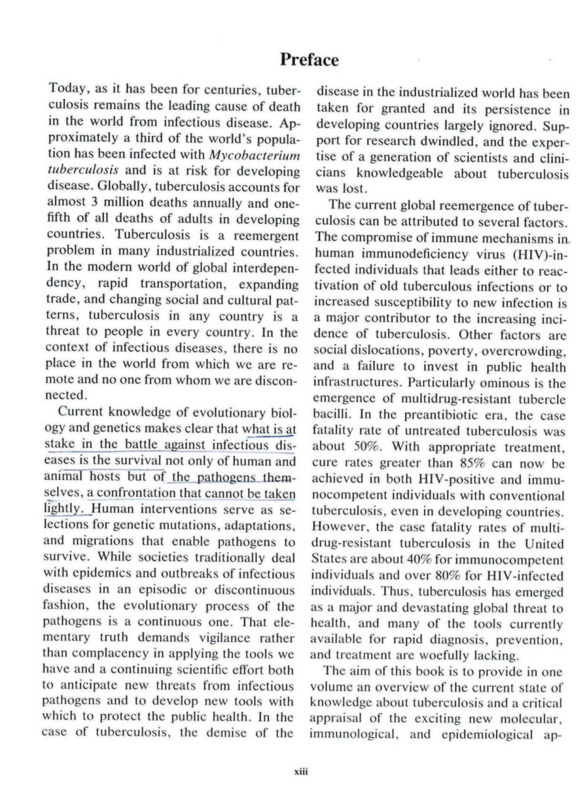
Impact of the HIV-AIDS Epidemic
of these patients lived in sub-Saharan Af
The pandemic of human immunodefi rica (Fig. 1). HIV seroprevalence rates of
ciency virus (HIV) infection and the evi more than 40% are common among patients
dence of an association between tuberculo with IB in many African countries. The
sis and HIV infection has caused marked annual risk of progression to active TB
increases in the incidence of TB in some among individuals infected with both HIV
countries. Because of its ability to destroy an^TBJs_5 to 10% (Narain et al., 1992).
the immune system, HIV has emerged as
I he result of this increased risk is evident
t le most significant risk factor for progres from the reported numbers of TB cases in
sion of dormant TB infection to clinical several countries. After years of declining
disease (Selwyn et al., 1989).
incidence of the disease, the number of
I he Global Programme on AIDS of the reported cases of TB increased dramati
WHO estimated that in 1992 at least 13 cally during the 1980s in many countries in
million adults and 1 million childZen had^ sub-Saharan Africa. Within a 7-year period
been infected with HIV worldwide (World from 1985 to 1991, the annual number of
Health Organization, 1993). Nearly 85% of
cases in Zambia nearly tripled, that in
HIV infections have occurred in developing Malawi more than doubled, and those in
countries, and the vast majority have oc Tanzania and Burundi increased by about
curred in the age group 15 to 49 years.
70 and 40%, respectively. The numbers of
It is estimated that about 1,700 million
deaths from TB have also increased in
peop e are infected with M. tuberculosis these countries (Narain et al., 1992).
(Kochi, 1991). The impact of HIV infection
The situation in some countries in South
on the TB situation is greatest in those
east Asia and the western Pacific is now
populations where the prevalence of TB
similar to that in Africa several years ago.
infection in young adults (who are at great
Since almost two-thirds of the world’s TBest risk of HIV infection) is high. By using
infected population is in Asia, entiy of HIV
estimates of the prevalence of TB infection
into Asian communities may result in large
in various regions, it has been estimated
increases
in HIV-associated TB in the com
that since the beginning of the HIV pan
ing years. In Bombay, HIV seroprevalence
6
Snider et al.
.s
Wk.
11 000
60 000
110 000+
9 000 +
17 000 +
•.?
3 760 000+
450 000
Figure 1. Estimated global distribution of adults who have been infected with HIV and TB, as of mid-1993.
Source of data is WHO Tuberculosis Programme.
among TB patients increased from 2% in mission to both HIV-positive and HIV
1988 to 10 to 15% in 1991 and 1992. In negative populations. Nevertheless, pre
northern Thailand, HIV seroprevalence in liminary data from the second round of the
TB patients increased from 5% in late 1989 National Tuberculin Survey in Tanzania,
to 15% in 1992, and there is evidence that where the TB control program has achieved
the incidence of TB is increasing (World an 80% cure rate of patients with newly
Health Organization, unpublished data).
diagnosed smear-positive cases during the
In many developing countries,
last 5 yearsTB
andhas
a 65% case detection rate, ________
emerged as the most common opportunistic suggest that the prevalence
infection iin
.__________of_________
disease associated with HIV infection. school children did not change appreciably
Twenty to forty-four percent of AIDS pa from 1983-1985 to 1989-1991, despite the
tients in Africa, 18% of patients in Haiti, increase in the. --------number of detected
---------- 1 new
and up to 25% of patients in some Latin smear-positive cases (Narain et al., 1992).
American countries, namely Brazil, Mex Thus, a good control program may be able i
ico, and Argentina, had clinical TB during to reduce to some extent the increased (
the course of HIV infection (Narain et al., chance of transmission.
1992).
HIV-infected patients with TB also have
It is still unclear how the increasing num a higher incidence of noninfectious extrabers of HIV-positive patients with TB will pulmonary forms of disease and higher
affect the transmission of TB in the commu mortality rates and thus may have a lesser
nity. It is possible that the increase in the impact on transmission than non-HIV-innumber of TB cases will increase TB trans fccted TB patients.
Chapter 1
TUBERCULOSIS IN INDUSTRIALIZED
COUNTRIES
United States
50
45 *
40
e and H1Vtheless, pre
round of the
in Tanzania,
has achieved
with newly
s during the
tection rate,
infection in
appreciably
despite the
itected new
al., 1992).
may be able
e increased
B also have
tious extraand higher
ave a lesser
lon-HIV-in-
• Observed cases
---- Expected cases
8> 30-
From 1953, when national surveillance
began, through 1984, the United States ex - 20 perienced a significant decline in TB cases:
15
51.700 Excess Casos
from 84,304 cases in 1953 to only 22,225
cases in 1984. The average annual decline
10
in cases was about 5.3% per year. From
00 81 82 83 &4 85 86 87 8S 89 90 91 92
1985 to 1992, however, the number of re
Year
ported cases has increased by about 20%. Figure 2. Numbers of expected and obsetved cases of
Using the trend for 1980 to 1984 to calculate 1 B in the United States from 1980 through 1992. Table
the number of expected cases, the Centers is from the CDC (Centers for Disease Control and
Prevention, 1993).
for Disease Control and Prevention (CDC)
estimates that from 1985 through 1992,
about 51,000 excess cases have accumu- I
largest increases in reported TB case numlated (Fig. 2). Case rates in urbani areas bers have occurred i
------------ in geographic areas and
have increased more rapidly than those ir.
in age groups heavily impacted by the HIV
rural areas.
epidemic. TB prevalence among AIDS pa
Of the 26,673 TB cases reported in 1992,
tients is high. Matching of TB and AIDS
71% occurred in racial and_ethnic minori
registries through 1990 revealed that 4.9%
ties. From 1985 to 1992, TB case numbers
oi reported AIDS cases were also in the TB
declined about 10% among non-Hispanic
registry (CDC, unpublished data). CDC
whites and 23% among Native Americans.
HIV seroprevalence surveys in TB clinics
However, case numbers increased 27%
have shown a high prevalence of HIV in
among blacks, 46% among Asians and Pa fection among TB patients. Pooled sero
cific Islanders, and 75% among Hispanics
prevalence data from 13 cities that tested at
From 1985 through 1992, all age groups,
least 50 serum samples per year for 1989,
except that of patients 65 years of age and
1990, and 1991 showed seroprevalence
older, experienced an increase in number
rates of 13.1% in 1989, 17.8% in 1990, and
of cases. The largest numerical and per
21.4% in 1991. Trend analysis from 1989
centage increases (+3,686; +55%) were through 1991 also showed significant up
among persons 25 to 44 years of age. How
ward trends in HIV seroprevalence among
ever, there was a 36% increase in case black, Hispanic, and white males with TB
numbers among 0- to 4-year-old patients
or suspected TB. Among females, the up
and a 34% increase among children 5 to 14
ward
trend was significant only for blacks
years old.
(Onorato et al., 1993).
Of all patients with TB reported to the
The United States has also experienced
CDC in 1992, 27% were born in another
an increasing number of outbreaks of TB.
country. The numbers and percentages of Outbreaks have occurred in a variety of
foreign-born patients increased from
settings, including hospitals, correctional
4,925 and 22% in 1986 to 7,270 and 27% in
facilities,
shelters for the homeless, resi
1992.
dential care facilities for patients with
In addition to the effect of immigration on
AIDS, nursing homes, and even crack
the change in the TB morbidity trend, there houses.
is evidence that the HIV epidemic is at least
The most serious problem has been the
in part contributing to this change. The
outbreaks of multidrug-resistant TB (MDR-
I
as of mid-1993.
7
I 35’
125
5 000 +
’
• Global Burden of Tuberculosis
8
I
Snider el al.
TB), i.e., outbreaks due to organisms resis■ ing the biennium 1988 through 1989 had TB
tant to isoniazid and rifampin (and ofteni (Raviglione et al., 1993).
other drugs as well). From 1990 through
1
In Australia, death rates have remained
1992, the CDC investigated outbreaks of stable at about 0.4/100,000, and notification
MDR-TB in eight hospitals and a state
rates of all cases have slightly increased
correctional system. As of November 1992, from 5.6/100,000 in 1986 to 5.9/100,000 in
297 cases of MDR-TB had been identified in 1990 (Cheah, 1992a). The HIV epidemic
these outbreaks. Most but not all of the has had little impact on the TB situation in
cases occurred in persons infected with Australia. Two-thirds of the new patients
HIV. Mortality was high (about 70%), and
reported in 1991 were foreign born (Cheah
the median interval from TB diagnosis to 1992b).
death was short (4 to 16 weeks). The out
In Canada, a similar stagnation of notifi
break investigations demonstrated trans
cations and rates has been observed over
mission of infection to health care workers. the past 6 years. In 1991, 2,044 cases were
At least 17 health care and correctional
reported (rate, 7.6/100,000). Foreign-born
workers have developed active TB with patients constituted 48% of all patients in
multidrug-resistant organisms (CDC, un 1989, and native Canadians constituted
published data).
20% of patients. TB mortality rates were
Factors contributing to nosocomial out-, stable at around 0.5/100,000 during recent
breaks include the convergence of highly
years (WHO, unpublished data [from the
susceptible, immunocompromised patients Canadian Centre for Health Information,
and TB patients; the delayed recognition of Ottawa]).
TB (because of unconsidered diagnoses,
In Japan, the downward trend of TB
nonclassical radiographic findings, and lab notifications continues. The average de
oratory delays); the delayed recognition of cline between 1980 and 1991 has been 3.5%
drug resistance; and the delayed initiation per year. However, this decline is smaller
of effective anti-TB treatment. Other fac than that seen in previous years. Further
tors contributing to nosocomial transmis more, the incidence of sputum-smear-posision include delayed initiation of isolation, tive cases has steadily increased since 1980.
inadequate ventilation for acid-fast bacillus
In general, mortality rates have regularly
(AFB) isolation, lapses in maintaining AFB decreased at about 4.6% per year since
isolation, inadequate duration of AFB iso 1980 (WHO, unpublished data [from the
lation, and inadequate precautions during
Japan Anti-Tuberculosis Association, To
cough-inducing procedures.
kyo]).
In New Zealand, case notifications have
recently increased from a nadir in 1988 of
Tuberculosis in Other Industrialized
295 cases reported. In 1991, 335 cases were
Countries
reported (rate, 9.9/100,000). Mortality rates
have decreased from 0.9 to 0.5/100,000 dur
Seven of 15 Western European countries ing the period 1980 to 1990 (WHO, unpub
(Denmark, Ireland, Italy, Netherlands, lished data [from the New Zealand Depart
Norway, Spain, and Switzerland) have also ment of Health, Wellington]).
recently experienced increases in reported
In Israel, TB notifications, after being /
cases (Raviglione et al., 1993). The major stable in the 1980s, recently increased to
factor responsible for most of these in 505 in 1991 (rate, 10.2/100,000). However,
creases appears to be immigration from if rates for Ethiopian immigrants are ex
higher-prevalence countries. However, in cluded from the data, the rate among native
Italy, 11.4% of AIDS patients reported dur- Jews in 1991 was 4.6/100,000 and that
I
1 1989 had TB
ave remained
d notification
tly increased
'.9/100,000 in
slIV epidemic
B situation in
new patients
born (Cheah,
■tion of notifibserved over
4 cases were
Foreign-born
11 patients in
constituted
J rates were
uring recent
ta [from the
Information,
rend of TB
average debeen 3.5%
is smaller
s. Furthersmear-posi1 since 1980.
/e regularly
year since
i [from the
nation, Toations have
• in 1988 of
cases were
rtality rates
00,000 durIO, unpubnd Dcpartifter being ,
creased to
However,
ts are exong native
and that
Chapter 1
among native non-Jews was 3.7/100,000.
Ethiopians, who generally constitute less
than 25% of all cases, constituted 50% of all
cases in 1985 and 55% of all cases in 1991,
following two waves of migration (Opera
tion Moses and Operation Shlomo) (WHO,
unpublished data [from the Israel Ministry
of Health, Jerusalem]).
In Turkey, TB notifications during the
past few years have decreased, and in 1990,
24,468 cases were reported (rate, 43.8/
100,000) (WHO, unpublished data [from the
Turkish Ministry of Health, Ankara]).
DRUG RESISTANCE
•
Global Burden of Tuberculosis
9
successfully introducing rifampin-containing short-course chemotherapy. Thus, the
incidence of acquired resistance was sub
stantially reduced, and the incidence of
primary resistance remains relatively low.
In many developing countries, particularly
in Asia, the incidence of acquired resis
tance remains high and the incidence of
primary resistance is higher than that in
industrialized countries, because national
TB control programs in the developing
countries have not been able to achieve a
high cure rate over a very long period, even
after the introduction of short-course che
motherapy.
This already serious situation may
quickly worsen as f
the HIV epidemic
spreads. The HIV epidemic may produce
increased levels of both acquired and pri
mary resistance not only by overtaxing the
national TB control programs as a result of
increased caseload but also by affecting
compromised immunity (Kochi et al
1993).
Acquired resistance is defined as resis
tance to at least one anti-TB drug that arises
during or after the course of treatment,
usually as a iresult of nonadhergnse to the
recommended regimen or of faulty pre
scribing. A high level of this type of resis
tance is a mark of a poorly functioning TB
control program.
Primary resistance is defined as the pres
ence of drug resistance to at least one
FUTURE TRENDS
anti-TB drug in a TB patient who has never
received prior treatment. It is caused by
Without recognition of the TB crisis con
infection with drug-resistant specimens fronting the world and prompt, effective
from another patient excreting a drug-resis action, the TB epidemic can be expected to
tant organism; many of these patients ac worsen for several reasons.
quired resistance as a result of inadequate
First, demographic forces are at work.
treatment. Thus, primary resistance is an Children born in past decades in regions
indicator of efficacy of TB control efforts in with high population growth rates are now
the past (Weyer and Kleeberg, 1992).
reaching the ages at which morbidity and
An accurate picture of the drug resis mortality for TB are high. Even if the
tance problem in the world is not available, age-specific rates of new cases do not in
because only a limited number of countries
crease, the changing sizes of the population
(both industrialized and developing) have a age groups will now begin to cause a large
reliable drug resistance surveillance sys increase in the number of TB deaths and
tem. However, limited available informa new cases.
tion indicates that, generally speaking,
Second, famine, war, and natural disasters
many industrialized countries that faced that create large populations of displaced,
severe MDR-TB in f
th,C ‘ atf J950s and early malnourished people in crowded living con1960s
successfully
reduced
the
rather short tim^ > •
• problem
‘
’ in
*.n aa ditions may cause increases in TB case rates,
of the sam ^reZ Y imPrOVm8
Third, age-specific TB incidence rates
of the same regimen used prevmusly and by can be expected to rise in those areas of the
10
Snider et al.
world where immunity of the population is
seriously challenged by HIV infection. By
mid-1993, the global cumulative number of
persons coinfected with HIV and tubercle
bacilli since the beginning of the HIV pan
demic was estimated to be over 5 million
(Fig. 1). In addition, HIV seroprevalence
among TB patients is expected to increase
further in areas like sub-Saharan Africa and
to increase at least threefold in areas like
Southeast Asia during the next decade. As
a result, while about 315,000 persons are
estimated to have developed HIV-associ
ated TB in 1990, more than 700,000 people
are expected to develop HIV-related TB in
1995. In the year 2000, the figure may reach
1.4 million. In 1990, 4.2% of all TB cases
were associated with HIV; in the year 2000,
an estimated 13.8% of all TB cases may be
associated with HIV (Dolin et al., in press).
In addition, there is the threat of the in
creasing incidence of drug-resistant strains.
This phenomenon is largely a consequence
of poorly managed and inappropriately fo
cused TB programs and is accelerated and
amplified by the HIV coinfection epidemic.
Drug-resistant strains are as contagious as
the normal TB bacillus. The cure rates of at
least 95% that can be achieved for regular
TB fall to 70% or less when isoniazid and
rifampin resistance occurs.
If the effectiveness and availability of TB
control do not improve substantially over
those existing now, more than 30 million
TB deaths and 90 million new cases are
expected to occur in the last decade of this
century. Conservative estimates indicate
that the incidence of TB can be expected to
increase to 8.8 million cases annually by
1995, 10.2 million cases annually by the
year 2000, and 11.9 million cases annually
by 2005 (Dolin et al., in press). Demo
graphic factors will account for three-quar
ters of the predicted increase in new cases.
Assuming that the availability and effec
tiveness of treatment programs remain at
the 1990 level, 3 million TB deaths can be
expected to occur annually by 1995, and 3.5
million deaths will be occurring annually by
the year 2000. Action must be taken now to
avert this global health disaster.
REFERENCES
Cauthen, G. M., and H. G. Ten Dam. 1988. Annual
risk of tuberculosis infection. Geneva'. W.H.OJTB
88:154.
Centers for Disease Control and Prevention. 1993.
Tuberculosis morbidity—United States, 1992. Mor
bid. Mortal. Weekly Rep. 42:696-704.
Cheah, D. 1992a. Tuberculosis notification rates, Aus
tralia final data for 1986 to 1990. Communicable Dis.
Intell. 16:234-236.
Cheah, D. 1992b. Tuberculosis notification rates, Aus
tralia, 1991. Communicable Dis. Intell. 16:398-400.
Dolin, P. J., M. C. Raviglione, and A. Kochi. Global
tuberculosis incidence and mortality during 19902000. Bull. W.H.O., in press.
Kochi, A. 1991. The global tuberculosis situation and
the new control strategy of the World Health Orga
nization. Tubercle 72:1-6.
Kochi, A., B. Vareldzis, and K. Styblo. 1993. Multidrug-resistant tuberculosis and its control. Res. Mi
crobiol. 2:104-110.
Murray, C. J. L., K. Styblo, and A. Rouillon. 1990.
Tuberculosis in developing countries: burden, inter
vention and cost. Bull. Int. Union Tuberc. Lung Dis.
65:6-24.
Narain, J. P., M. C. Raviglione, and A. Kochi. 1992.
HIV-associated tuberculosis in developing coun
tries: epidemiology and strategies for prevention.
Tuberc. Lung Dis. 73:311-321.
Onorato, I., S. McCombs, M. Morgan, and E. McCray.
1993. HIV infection in patients attending tuberculo
sis clinics. United States, 1988-1992. Program Abstr. 33rd Intersci. Conf. Antimicrob. Agents
Chemother., abstr. 1363.
Raviglione, M. C., P. Sudre, H. L. Rieder, S. Spinaci,
and A. Kochi. 1993. Secular trends of tuberculosis in
Western Europe. Bull. W.H.O. 71:297-306.
Selwyn, P. A., D. Hartel, and I. A. Lewis. 1989. A
prospective study of the risk of tuberculosis among
intravenous drug abusers with human immunodefi
ciency virus infection. N. Engl. J. Med. 320:545550.
Styblo, K., J. Meiger, and 1. Sutherland. 1969. The
transmission of tubercle bacilli, its trend in a human
population. Bull. Int. Union. Tuberc. Lung Dis.
42:5-104.
Styblo, K., and A. Rouillon. 1981. Estimated global
incidence of smear-positive pulmonary tuberculosis.
Unreliability of officially reported figures on tubercu-
Chapter 1
?5, and 3.5
nnually by
<en now to
1988. Annual
: W.H.OJTB
ention. 1993.
s, 1992. Mor-
>n rates, Ausunicable Dis.
•n rates, Aus. 16:398-400.
<ochi. Global
during 1990situation and
Health Orga-
1993. Multirol. Res. Mimilion. 1990.
mrden, interrc. Lung Dis.
Kochi. 1992.
doping coun• prevention.
d E. McCray,
ig tuberculoProgram Ab-ob. Agents
r, S. Spinaci,
iberculosis in
-306.
wis. 1989. A
jlosis among
immunodefied. 320:5451. 1969. The
d in a human
. Lung Dis.
nated global
tuberculosis,
s on tubercu-
losis. Bull. Int. Union Tuberc. Lung Dis. 56:118-126.
Sutherland, I. 1976. Recent studies in the epidemiol
ogy of tuberculosis, based on the risk of being
infected with tubercle bacilli. Adv. Tuberc. Res.
19:1-63.
Weyer, K., and H. H. Kleeberg. 1992. Primary and
acquired drug resistance in adult black patients with
tuberculosis in South Africa: results of a continuous
• Global Burden of Tuberculosis
11
national drug surveillance programme involvement.
Tuberc. Lung Dis. 73:106-112.
World Health Organization. 1989. Childhood tubercu
losis and BCG vaccine. In EPI Update Supplement.
World Health Organization, Geneva.
World Health Organization. 1993. Global Programme
on AIDS. The HIV/AIDS pandemic: 1993 overview.
WHO/EPA/CNP/EV A/93.1.
Tuberculosis: Pathogenesis. Protection, and Control
Edited by Barry R. Bloom
<> 1994 American Society for Microbiology. Washington. DC 20005
rculosis. Har
tass.
is of Disease,
g, Springfield,
Courting Danennis, Golden
ving. St. Mar-
Chapter 3
Overview of Clinical Tuberculosis
ks of disease
a simulation
7:483^196.
. Ucko. 1964.
Rev. Respir.
Philip C. Hopewell
Evidence for
n. JAMA 241:
The clinical expression of infection with
Mycobacterium tuberculosis is quite varied
and depends on a number of identified
factors. Table 1 lists both host- and mi
crobe-related characteristics as well as the
consequences of their interactions that in
fluence the manifestations of tuberculous
infection. Among generally healthy per
sons, infection with M. tuberculosis is
highly likely to be asymptomatic. Data
from a variety of sources suggest that the
lifetime risk of developing clinically evident
tuberculosis after being infected is approx
imately 10%, with a 90% likelihood of the
infection remaining latent (Comstock,
1982). Only a positive tuberculin skin test
indicates the presence of the organism in
persons with latent infections. In specific
subpopulations, for example, in persons
with immunodeficiency states or in infants,
the proportions who develop evident tuber
culosis are much higher (Allen et al., 1992;
Comstock, 1982; Selwyn et al., 1992).
Immunization with bacillus of Calmette
and Guerin (BCG) in persons with intact
cell-mediated immunity minimizes the risk
of early disseminated tuberculosis, espe
cially in children. In addition to host fac-
<w, and W. H.
of streptomy. Tuberc. 71:
:ako: Estudio
ercolombinas
ibro. La Paz,
Discussion of
■enic PhysiolC.V. Mosby
ohn Harvard.
U. Sultan, F.
ifectiousness
ziolet irradiactiousness of
Dis. 85:5112. Hydrazine
on, Marsilid)
iseous-pneu65:402-428.
y of the dis2. Bull. Int.
almette and
ace to tuber-
nimal tuberuberculosis.
yHoeber Inc.,
I
ice of tubcr195-225.
i
Philip C. Hopewell • University of California, San
Francisco, and Division of Pulmonary and Critical
Care Medicine, San Francisco General Hospital, 1001
Potrero Avenue, Room 5K1, San Francisco, Califor
nia 94110.
25
tors, there probably are factors related to
the organism itself, such as its virulence or
predilection for specific tissues, that influ
ence the outcome and features of the infec
tion; however, these features of the organ
ism have not been characterized.
The most obvious and important factor
influencing the clinical features of tubercu
losis is the site of involvement. Prior to the
beginning of the epidemic of infection with
the HIV, approximately 85% of reported
tuberculosis cases were limited to the
lungs, with the remaining 15% involving
only nonpulmonary sites or both pulmo
nary and nonpulmonary sites (Farer et al.,
1979) (Fig. 1). This proportional distribu
tion is substantially different among per
sons with HIV infection. Although there
are no national data that describe the sites
of involvement in HIV-infected persons
with tuberculosis, in one large retrospec
tive study of tuberculosis in patients with
advanced HIV infection, it was reported
that 38% had only pulmonary involvement,
30% had extrapulmonary sites, and 32%
had both pulmonary and nonpulmonary in
volvement (Small et al., 1991). The multi
plicity of sites in HIV-infected persons is
typical of what is seen in an individual
having an immune system that is limited
in its ability to contain infection with M.
tuberculosis. Included in this category are
infants, the elderly, and persons with pri-
26
Hopewell
77-7cltntea. features of luberculosis
Host factors
--- ------------------------Microbial factors
Age
Host-microbe interaction
Virulence
of organism (?)
Immune status
Sites of involvement
Predilection (tropism) for
Specific immunodeficiency states
Severity of disease
specific
tissues
(?)
Malnutrition
Genetic factors (?)
Coexisting diseases
Immunizadon with bacillus of Calmette and
Guerin (BCG)
KX-j K-5
SYS'] EMIC AND REMOTE EFFECTS
1
OF
rLhe frequency with which fever has been
observed in patients with tuberculosis var-
tuberculosis
Tuberculosis occurring at any site may
produce symptoms and findings that are no^
specifically related to the organ or tissue
mvo ved but; „
are systcm.c na(sue
r are remote from the site of disease
Systemic manifestations of the disease, inSv 8meder; h3', 3156’ a”d Weight ,oss> are
kely mediated by cytokines, especially
^umor necrosis factor alpha (TNF-a). Expenmental data suggest that TNF-a is an
et'al wTX1™'6'7 37 t0 80% (A'a'’S'>
bt Kihi 8’&
1 et aI-’1981j-In a study ■
198U
h and,Coworkers (Kiblawi et ai.,
c,9fi8e I Wh'Ch th£ feVer resP°nse was spe
cifically examtned, 21% of patients had no
fever at any point in the course of hospitaltUberCulosis- Of 'he febrile pahents, 34% were afebrile within 1 week and
64% were afebrile within 2 weeks. The
median duration of fever after beginning
eatment was 10 days, with a range of 1 to
100
(Extra-
90 _
pulmonary
Il4.6%
Bone/Joint 8.5%
80 -
Other 9.3%
70 -
o
5
Peritoneal 3.7%
Meningeal 4.2%
60 -
Miliary 9.8%
50 -
Genitourinary 16.0%
40 -
Pleural 21.5%
30 _
20 -
10 -
■■Pulmonary
Lymphatic 27.0%
0
ALL CASES
ALL
EXTRAPULMONARY
CASES
Hgure I. Distribution of sites of involvement in
newly reported cases of tuberculosis fa 1978 prior to the
epidemic of infection with HIV
a
a
Chapter 3
abe interaction
✓olvement
f disease
smic effects
al., 1990;
ystemic efquantified.
:r has been
culosis var)% (Arango
. In a study
•lawi et al.,
>e was spe•nts had no
of hospitalfebrile pa1 week and
'eeks. The
beginning
nge of 1 to
i
prior to the
109 days. Weight loss, weakness, and mal
aise appear to be less common but are more
difficult to quantify.
In addition to these generalized effects of
tuberculosis, there are remote manifesta
tions that are not a result of the anatomic
site of involvement. These include hemato
logic abnormalities, hyponatremia, and psy
chological disorders. The most common
hematologic manifestations of tuberculosis
are increases in the peripheral blood leukocyte count and anemia, each of which oc
curs in approximately 10% of patients with
apparently localized tuberculosis (Cam
eron, 1974; Carr et al., 1964). The increase
in leukocyte counts is usually slight, but
leukemoid reactions may occur. Leukope
nia has also been reported. An increase in
the peripheral blood monocyte and eosino
phil counts also may occur with tuberculo
sis. Anemia is common when the infection
is disseminated. In some instances, anemia
or pancytopenia may result from direct
involvement of the bone marrow and thus
be a local rather than a remote effect.
Other than weight loss, the most frequent
metabolic effect of tuberculosis is hy
ponatremia, which in one series was found
to occur in 11% of patients (Chung and
Hubbard, 1969). Hyponatremia is caused
by production of an antidiuretic hormonelike substance found within affected lung
tissue (Vorken et al., 1970). The poor prog
nosis that in the prechemotherapy era was
associated with hyponatremia was proba
bly related simply to the amount of lung
involved and perhaps to adrenal involve
ment. In addition, because the syndrome of
inappropriate secretion of antidiuretic hor
mone is also associated with central ner
vous system disorders, hyponatremia may
be a feature of central nervous system
tuberculosis.
The psychological effects of tuberculosis
are very poorly defined but were commonly
recognized prior to the advent of effective
therapy. These effects include depression
and, on occasion, hypomania. The best
• Overview of Clinical Tuberculosis
27
descriptions of the psychological alter
ations in patients with tuberculosis are
found in literary works, such as Thomas
Mann’s The Magic Mountain, rather than
in medical writings.
In many patients, tuberculosis is associ
ated with other serious disorders, including
HIV infection, alcoholism, chronic renal
failure, diabetes mellitus, neoplastic dis
eases, and drug abuse, to name but a few.
The signs and symptoms of these diseases
and their complications can easily obscure
or modify those of tuberculosis and can
result in considerable delays in diagnosis or
in misdiagnoses for extended periods, espe
cially in patients with HIV infection
(Kramer et al., 1990). For this reason it is
important that clinicians have an under
standing of the diseases with which tuber
culosis may coexist and have a high index
of suspicion for a combination of the two
disorders.
TUBERCULIN SKIN TESTING
As noted above, a positive tuberculin
skin test is usually the only evidence of
latent tuberculous infection (Sbarbaro,
1986). Among persons with symptoms or
clinical findings of tuberculosis, the tuber
culin skin test may provide useful diagnos
tic information. However, in an individual
patient a positive test (usually defined as an
induration of >10 mm in immunocompetent
persons and >5 mm in persons with HIV
infection) does not establish a diagnosis and
a negative test does not exclude tuberculo
sis. Up to 25% of apparently immunocompetent persons will have negative tubercu
lin skin tests at the time of diagnosis of
tuberculosis (Nash and Douglas, 1980).
Among patients with tuberculosis and HIV
infection, the frequency of positive tuber
culin reactions varies considerably depend
ing on the degree of immune compromise
(Reider et al., 1989).
30
Hopewell
I
endogenous reactivation of latent infection
material may be spread via the airways into
usually causes abnormalities in the upper the lower portions of the lung or to the
lobes of one or both lungs. Cavitation (de
other lung. Erosion of a parenchymal focus
struction of lung tissue) is common in this of tuberculosis into a blood or lymph ves
form of tuberculosis. The most frequent
sel may result in dissemination of the or
sites of involvement are the apical and
ganism and a “miliary” (evenly distributed
posterior segments of the right upper lobe small nodules) pattern on the chest film
and the apical-posterior segment of the left (Fig. 5).
upper lobe (Fig. 4). Healing of the tubercu
In patients with HIV infection, the nature
lous lesions usually results in development
of the radiographic findings depends to a
of a scar with loss of lung parenchymal
certain extent on the degree of immuno
volume and, often, calcification. In the im
compromise produced by the infection. Tu
munocompetent adult with tuberculosis, in- berculosis that occurs relatively early in the
trathoracic adenopathy is uncommon but
course of HIV infection tends to have the
may occur, especially with primary infec typical radiographic findings described
tion. As tuberculosis progresses, infected above (Chaisson et al., 1987; Pitchenik and
Chapter 3
I
•
Overview of Clinical Tuberculosis
'4^1®
!
Figure 5. Portion of chest radiograph showing nodular lesions in a
uberculosis in
patient with disseminated tuberculosis.
!
Rubinson, 1985). With more advanced HIV
disease, the radiographic findings become
more “atypical”: cavitation is uncommon
and lower lung zone or diffuse infiltrates
and intrathoracic adenopathy are frequent
(Fig- 6).
M
rways into
or to the
/mal focus
ymph vesof the oriistributed
-best film
Bacteriologic Evaluation
the nature
"nds to a
immuno
bion. Tu•rly in the
have the
iescribed
tenik and
I
i
At |----- a definitive diagnosis of tupresent,
berculosis
---- 3 can be established only by isola
tion of tubercle bacilli in culture, although
tests that identify specific M. tuberculosis
ONA should soon be available for clinical
use-.When the lung is involved, sputum is
the initial diagnostic specimen of choice.
Sputum specimens should be collected at
the time of the initial evaluation. Single
early-morning specimens have aa higher
yield and a lower rate of contamination
than pooled specimens. The sensitivity of
sputum examination increases with the
number of specimens, but there is no in
crease in cumulative recovery of organisms
with more than five specimens, and the
increased yield between three and five
specimens is slight (Kubica et al., 1975)
There are several ways of obtaining spec
imens from patients who are not producing
sputum. The first and most useful is induc
ing sputum production by the inhalation of
a mist of hypertonic (3 to 5%) saline gener
ated by an ultrasonic nebulizer. This is a
benign and well-tolerated procedure, al-
31
K
32
Hopewell
I;
fflvTnLtfon"'31 VieW’ CheS' rad‘Ograph’ showing diffuse infiltration caused by M.
tuberculosis in a patient with
though bronchospasm may occasionally be (Burk et al., 1978; Danek and Bower, 1979;
precipitated in asthmatics. Samples of gas So et al., 1982). For larger nodular lesions,
tric contents obtained via a nasogastric
needle aspiration biopsy may also provide
tube have lower yields than induced spu specimens from which M. tuberculosis can
tum, and the procedure is more compli be isolated. This technique is more suited
cated and uncomfortable for the patient. to the evaluation of lesions when there is a
However, in children and some adults, gas suspicion of malignancy.
tric contents may be the only specimen that
In some situations, a therapeutic trial of
can be obtained.
antituberculosis chemotherapy may be in
Usually, fiberoptic bronchoscopy is the dicated before more invasive studies are
next diagnostic step if the sputum is nega undertaken (Gordin et al., 1989). Im
tive or cannot be obtained by induction.
provement in the chest film concomitant
In general, the bronchoscopic j
___ 2_._
procedure
with antituberculosis treatment would be
should include bronchoalveolar lavage and sufficient reason for making a diagnosis of
transbronchial lung biopsy. The yield of tuberculosis and continuing with a full
bronchoscopy has been high both for mili course of therapy. If a response is going
ary tuberculosis and for localized disease to occur, it should be seen within 3
Chapter 3
1-.
I
-*- Respiratory TB Other than Lung
-e- Genitourinary System
0.8-
I
Overview of Clinical Tuberculosis
-e- Meninges or Central Nervous System
>
/
•
tr Disseminated
— Lymphatic System
Zx
Case Rate o.eper
100,000
population 0.4-
0.2i
•o
0
o—
0-4
I
1
5-14
15-24
i
25-34
i---------- 1
1
T------------1
35-44 45-54 55-64
65+
Age
Figure 7. Age-specific case rates for the most frequent forms of extrapulmonary tuberculosis.
I
in a patient with
I
Bower, 1979;
'ular lesions,
also provide
rculosis can
more suited
3n there is a
mtic trial of
' may be in
studies are
1989). Imancomitant
t would be
iagnosis of
vith a full
’6 is going
within 3
months of starting treatment. In the
United States, the criteria for defining a
case of tuberculosis allow for culture neg
ativity if the patient in question has a
positive tuberculin skin test and responds
to multidrug chemotherapy.
extrapulmonary tuberculosis
I
l
As noted above, prior to the epidemic of
more of a diagnostic and therapeutic prob
lem than pulmonary tuberculosis. In part,
this problem relates to its being less com
mon and therefore less familiar to most
clinicians (Alvarez and McCabe, 1984Weir and Thornton, 1985). In addition, ex
trapulmonary tuberculosis involves rela
tively inaccessible sites, and because of the
nature of the sites involved, fewer bacilli
reported cases of tuberculosis involved
inaccessible sites makes bacteriologic con
only extrapulmonary sites (Farer et al.,
firmation of a diagnosis more difficult, and
1979). For reasons that are not understood,
invasive procedures are frequently required
as rates of pulmonaiy tuberculosis de
to establish a diagnosis.
creased, rates of extrapulmonary disease
The relative frequencies of tuberculosis
remained constant, resulting in an increas
at various sites in persons without immuno
ing proportion of cases being extrapulmo
compromise are shown in Fig. 1, and dis
nary. With the onset of the HIV epidemic
tribution by age is shown in Fig. 7 (Farer et
however, both absolute and relative rates
al.,
1979). As can be seen, in general, the
of extrapulmonary involvement have in
incidence
for each extrapulmonary site in
creased.
creases
with
increasing age, except for lym
Extrapulmonary tuberculosis presents
phatic and meningeal tuberculosis, which
3.
34
Hopewell
Table 2, Recovery of A/, tuberculosis from various sites in patients with tuberculosis and HIV infection"
Specimen
Sputum
Bronchoalveolar lavage
Transbronchial biopsy
Lymph node
Blood
Bone marrow
Cerebrospinal fluid
Urine
Other2’
___________ No- Positive/n°- tested (%)
Smear
Culture
43/69 (62)
9/44 (20)
1/10 (10)
21/44 (48)
64/69 (93)
39/44 (89)
7/10 (7)
39/44 (91)
15/46 (33)
13/22 (62)
4/21 (19)
12/17 (71)
24/31 (76)
4/22 (18)
5/31 (16)
" Data are from Small et al. (1991).
Plonral An«z4
. .
i- i
Pleural
fluid or tissue, pericardial
fluid or tissue, liver peritoneal fluid, abscess drainage, or bone.
are relatively more common in young chil
dren.
I
I:
■
scriptive epidemiology of disseminated
tuberculosis. Disseminated or miliary tu
berculosis occurs because of the inade
Extrapulmonary Tuberculosis in
quacy of host defenses in containing tu
HIV-Infected Patients
berculous infection. This failure of
Presumably, the basis for the frequency containment may occur in either latent or
of extrapulmonary tuberculosis among pa recently acquired tuberculous infection.
tients with HIV infection is the failure of Because of HIV or other causes of immu
the immune response to contain M. tuber nosuppression, the organism proliferates
culosis, thereby enabling hematogenous and disseminates throughout the body.
dissemination and subsequent involvement Multiorgan involvement is probably much
of single or multiple nonpulmonary sites. more common than is recognized, because
As evidence of this sequence, tuberculosis generally, once M. tuberculosis is identified
bacillemia has been documented in HIV- in any specimen, other sites are not evalu
infected patients on a number of occasions ated. The term “miliary” is derived from
(Handwerger et al., 1987; Kramer et al., the visual similarity of the lesions to millet
1990; Shafer et al., 1989). Because of the
seeds. Grossly, these lesions are 1- to
frequency of extrapulmonary tuberculosis
2-mm-diameter yellowish nodules that, his
among HIV-infected patients, diagnostic
tologically, are granulomas. Persons with
specimens from any suspected site of dis
HIV infection may not be able to form
ease should be examined for mycobacteria.
granulomas; thus, the individual lesions
Moreover, cultures of blood and bone mar
may not be present. Instead, a diffuse uni
row may reveal M. tuberculosis in patients
form pattern of lymphocytic infiltration and
who do not have an obvious localized site
edema is seen.
of disease but who are being evaluated
Although disseminated tuberculosis near
because of fever. Table 2 lists the sites from
ly
always involves the lungs, it is consid
which M. tuberculosis was recovered in a
ered
among the extrapulmonary forms of
group of patients with advanced HIV infec
the
disease
because of the multiplicity of
tion (Small et al., 1991).
organs affected. In the past, miliary tuber
culosis occurred mainly in young children;
Disseminated Tuberculosis
currently, however, except among HIVThe epidemic of HIV infection has con infected persons, it is more common among
siderably altered the frequency and de- older persons (Barer et al., 1979). The shift
3
o
Chapter 3
’ infection"
m age-specific incidence presumably has
been caused by the paucity of new infec
tions relative to the number of endogenous
reactivations that take place in the United
States. The sex incidence is nearly equal
except in the HIV-infected population,
wherein men predominate.
Culture
64/69 (93)
39/44 (89)
7/10 (7)
39/44 (91)
15/46 (33)
13/22 (62)
4/21 (19)
12/17 (71)
24/31 (76)
35
bin and alanine aminotransferase levels
may also be increased.
I he chest film is abnormal in most but
not all patients with disseminated tuberculosis. In the series reported by Grieco and
Chmel (1974), only 14 (50%) of 28 patients
.. -r,.rak™nl
geminated
liliary tuie inade(ining tuilure of
latent or
nfection.
3f immu)liferates
e body.
»ly much
because
dentified
)t evalu
ed from
to millet
e 1- to
hat, his■ns with
to form
lesions
use uni
ion and
•is nearconsid>rms of
icity of
' tuberu’ldren;
: hivamong
ie shift
• Overview of Clinical Tuberculosis
1
-Seminated tuberculosis, the clinical had a miliary pattern. Overall, it appears
i amfestations are protean. The presenting that at the time of diagnosis, approximately
eifimp H S and 4SIgnS are generalIy nonsPe- 85% of Patients have the characteristic ra^^
c and are dominated by the systemic biographic findings of miliary tuberculosis
effects, particularly fever, weight loss, an- Other
°ther radiographic
radiographic abnormalities
abnormalities imay be
7neakness (Grieco and Chmel, P^sent
as
well.
These
include upper "lobe
lobe
present
19/4, Munt, 1971; Prout and Benatar, 1980; ^filtrates with or without cavitation pleuSahn and Neff, 1974; Slavin et al., 1980). ral
raJ effusion,
effusion, and
and pericardial
pericardial effusion,
effusion In
Other symptoms depend on the relative patients
Patients with HIV infection, tthe
’._ radio
severity of disease in the organs involved. graphic pattern is one of diffuse infiltration
Cough and shortness of breath are com rather than discrete nodules.
mon; headache and mental status changes
____is positive less
1 he tuberculin skin test
are less frequent and are usually associated frequently in patients with disseminated
with meningeal involvement (Munt, 1971). tuberculosis than in those with other forms
Physical findings are likewise variable. Fe of the disease. The rate of positivity at the
ver, wasting, hepatomegaly, pulmonary time of diagnosis in apparently immuno
findings, lymphadenopathy, and spleno competent persons ranges from approxi
megaly occur in descending order of fre mately 50 to 75% (Grieco and Chmel, 1974;
quency. The only physical finding that is Munt, 1971; Sahn and Neff, 1974; Slavin et
specific for disseminated tuberculosis is the al., 1980). As the disease is treated, tuber
choroidal tubercle, a granuloma located in culin reactivity tends to return unless there
is systemic immunocompromise.
the choroid of the retina (Massaro ct al.
1964).
Autopsy series have shown the liver,
Initial screening laboratory studies are lungs, bone marrow, kidneys, adrenals, and
not particularly helpful for the diagnosis of spleen to be the organs most frequently
miliary tuberculosis. Both leukopenia and involved in miliary tuberculosis, but any
leukocytosis may be seen, but the majority organ can be the site of disease (Slavin et
ol patients have normal leukocyte counts. aL, 1980). Because of the multiplicity of
Anemia is common and may be normo sites involved, there are many potential
cytic, normochronic, or microcytic and sources of material used to provide a diag
hypochromic. Coagulation disorders are nosis. Acid-fast smears of sputum are pos
unusual, but disseminated intravascular co itive in 20 to 25% of patients, and M.
agulation has been reported in association tuberculosis is isolated from sputum in 30
With
”a-
and Chmel>
with miliary tuberculosis in severdy
severely’ill ppa
bents (Huseby and Hudson, 1976; Murray sZnl ai Z c",
5’
pt
ui
imos
tt
---.
.
.
y
oiavin
et
al.,
1980).
Gastric
washings
or
ct al., 1978). Hyponatremia also occurs, as
induced
sputum
may
be
positive
when
the
discussed above. The most frequent abnor
mality of liver function is an increased patient is not expectorating spontaneously,
in a patient with an abnormal chest film and
alkaline phosphatase concentration. Bilirunegative sputum examinations, bronchos-
36
,1
Hopewell
copy should be the next step. Combinations
In non-HIV-infected persons with tuber
of bronchoalveolar lavage and transbronculous lymphadenitis, systemic symptoms
chcal biopsy would be expected to
to have
have a
. .............. ulllV
oc» uthere is concomiunless
.are. n°.t comnion
high yield. Other potential sites ‘
itcs lor biopsy tant tuberculosis elsewhere.
- -------'■ The frequency
include liver and bone marrow, each of
of
i
which has a high likelihood of showing ot pulmonary involvement in reported se
ries
nes
of
patients
with
tuberculous
lymphad
granulomas (70 to 80%) but only a 25 to 40%
from ap
enitis
is
quite
variable,
ranging
chance of providing bacteriologic confirma
proximately
5
to
70%.
In
HIV-infected
tion (Sahn and Neff, 1974). Urine is easy to
asso
commonly assoobtain and may be positive in up to 25% of persons, lymphadenitis is commonly
ciated with multiple-organ involvement, al
patients (Sahn and Neff, 1974). Selection of
though localized lymphadenitis, as de
other potential sources of diagnostic mate
scribed above.
above, may occur as well.
rial should be guided by specific findings.
The diagnosis of tuberculous lymphade
nopathy is established by lymph node
Lymph Node Tuberculosis
biopsy or aspiration with histologic examiHIV enidemir
.
nat,On’ incIuding stains
acid-fast organPrior to the I""
..?ilympL.Ilode ISkmS’ and culture of ‘he material. Smears
tuberculosis made up approximately 20% of
show acid-fast organisms in approximately
the cases of extrapulmonaiy tuberculosis in
25 to 50% of biopsy specimens, and M
the United States (Farer et al., 1979) A],
though the H-' ’
?.fItUbCrCU,°SiS applies to ^Phatic tuberlXShSatic----hturber
cruIoX°
* aare
two is^elat^more
main differences: tamT
is
---------common among cMdren relatively
and h ocmore fro™ ™m™ocompetent patients. In immuamong children, and it occurs
Pacific Islanders than among blacks and
whites. Among HIV-infected persons, the
demographic features of tuberculous lymph
Pleural Tuberculosis
adenitis parallel those of HIV infection.
I he epidemiology of pleural tuberculosis
luberculous lymphadenitis usually pre
parallels that of the overall pattern for tu
sents as painless swelling of one or more
berculosis, with the disease being more
lymph nodes. The nodes most commonly
common
among males and increasing in
involved are those of the posterior or ante
incidence with increasing age between ages
rior cervical chain or those in the supracla
5 and 45 (Farer et al., 1979). As noted
vicular fossa. Frequently, the process is
above,
this epidemiologic pattern is modi
bilateral, and other noncontiguous groups
fied by the occurrence of HIV infection,
of nodes can be involved (Kent, 1967). At
least initially, the nodes are discrete and the although pleural involvement seems rela
overlying skin is normal. With continuing tively less frequent among HIV-infected
persons.
disease, the nodes may become matted and
1 here are two mechanisms by which the
the overlying skin inflamed. Rupture of the
pleural space becomes involved in tubercu
node can result in formation of a sinus tract
losis, and the difference in pathogenesis
which may be difficult to heal. Intrathoracic
results
in different clinical presentations,
adenopathy may compress bronchi, caus
approaches
to diagnosis, treatment, and
ing atelectasis and leading to lung infection
sequelae.
Early
in the course of a tubercu
and perhaps bronchiectasis.
lous infection, a few organisms may gain
I
IJ
I
I
Chapter 3
svith tuber
symptoms
; concomifrequency
ported selymphadfrom apV-infected
□nly assoement, al, as deI.
ymphadeiph node
;ic examiist organl. Smears
Jximately
. and M.
y 70% of
tubercuig granu' samples
In immumay be
evsky et
irculosis
1 for tuig more
asing in
*en ages
s noted
s modifection,
ns relanfectcd
lich the
ubercugenesis
rations,
it, and
ibercuty gain
1
• Overview of Clinical Tuberculosis
37
access to the pleural space, and in the whom tuberculous pleuritis is ultimately
presence of cell-mediated immunity, they diagnosed (Levine et al., 1970; Scharer and
can cause a hypersensitivity response McClement, 1968). In a patient who has a
(Berger and Mejia, 1973; Ellner, 1978). pleural effusion that remains undiagnosed
Commonly, this form of tuberculous pleu after a full evaluation, including pleural
ritis goes unnoticed, and the process re biopsy, and who has a positive tuberculin
solves spontaneously. In some patients, skin test reaction, antituberculosis treat
however, tuberculous involvement of the ment should be initiated.
pleura is manifested as an acute illness with
The second variety of tuberculous in
fever and pleuritic pain. If the effusion is volvement of the pleura is a true empyema
large enough, dyspnea may occur, although (pus in the pleura). This condition is much
the effusions generally are small and rarely less common than tuberculous pleurisy
are bilateral. In approximately 30% of pa with effusion and results from a large num
tients, there is no radiographic evidence of ber of organisms spilling into the pleural
involvement of the lung parenchyma; how space, usually from rupture of a cavity or
ever, parenchymal disease is nearly always an adjacent parenchymal focus via a bronpresent, as evidenced by findings by lung chopleural fistula (Johnson et al., 1973). A
dissections (Stead et al., 1955).
tuberculous empyema is usually associated
The diagnosis of pleural tuberculosis is with evident pulmonary parenchymal dis- *
generally established by analysis of pleural ease on chest films, and air may be seen in
fluid and/or pleural biopsy. A thoracentesis the pleural space. In this situation, the fluid
(aspiration of fluid from the chest) should is generally thick and cloudy and may con
be performed, and sufficient fluid for cell tain cholesterol, causing the fluid to look
count, cytologic examination, biochemical like chyle (pseudochylous effusion). The
analysis, and microbiologic evaluation fluid is exudative and usually has a rela
should be obtained, leaving enough to allow tively high leukocyte count, with nearly all
a needle biopsy to be performed if the fluid of the leukocytes being lymphocytes. Acid
is exudative and no diagnosis is evident. fast smears and mycobacterial cultures are
The fluid is nearly always straw colored, usually positive, making pleural biopsy un
although it may be slightly bloody. Cell necessary. This type of pleural involvement
counts are usually in the range of 100 to has a tendency to burrow through soft tis
5,000/jjJ (Jay, 1985). Early in the course of sues and may drain spontaneously through
the process, polymorphonuclear leuko the chest wall. An example of this type of
cytes may predominate, but mononuclear tuberculosis is shown in Fig. 8.
cells soon become the majority. The fluid is
exudative, with a protein concentration
Genitourinary Tuberculosis
greater than 50% of the serum protein con
centration, and the glucose level may be
As with pleural tuberculosis, the epide
normal to low.
miologic pattern of genitourinary tubercu
Because few organisms are present in the losis parallels that of tuberculosis in general
pleural space, smears of pleural fluid are except that the incidence is nearly equal in
rarely positive, and M. tuberculosis is iso men and women. The pathogenesis appears
lated by culture in only 20 to 40% of pa to be one of seeding of the kidney at the
tients with proved tuberculous pleuritis time of the initial infection and bacillcmia.
(Levine et al., 1970; Scharer and McIn patients with genitourinary tuberculo
Clement, 1968). A single blind needle bi sis, local symptoms predominate, and sys
opsy of the pleura will confirm the diagno temic symptoms are less common (Chris
sis in approximately 65 to 75% of patients in
tensen, 1974; Simon el al., 1977). Dysuria,
38
Hopewell
120 I V
14O
Mh
LPG •FOV
10 0 Mhl
0 0 TILT
1£0 KV
120 Mh
LPG SFOV
5 0 MM
0 0 TILT
2 0 SEC 17 17 37
Chapter 4
•
Epidemiology of Tuberculosis
?e
f all tuberculo)bserved along
’erculosis inciFhis phenomeNew York. In
lent strains in
or more drugs
are dramatic
iberculosis in
-atin America
sle resistance
h AIDS and
ead of drug
ated to HIV
s. First, nons drug users
risk for HIV
R strains are
populations
'ith tubercu'e transmisive persons
/ in congretelescoping
neration of
/ infections
Ives be unnoncomplisk of HIV
and active
e likely to
>pulations.
with HIV
leveloping
ntial for a
h intraveigh backi as Thai-
epide; the bai
tion and
Jveloped
and par-
57
ticularly in Africa, coinfection with M. tu
in cohorts of HIV-infected and uninfected urban
berculosis and HIV threatens to double or
Rwandan women. Ain. Rev. Respir. Dis. 146-1439—
1444.
triple the clinical burden of tuberculosis.
De Cock notes that “with current re Andersen, S., and A. Geser. 1960. The distribution of
tuberculosis infection among households in African
sources, existing tuberculosis programs in
communities. Bull. W.H.O. 22:39-60.
sub-Saharan Africa will not cope with the Bermejo, A., H. Veeken, and A. Berra. 1992. Tuber
increased tuberculosis burden” (De Cock
culosis incidence in developing countries with higlx_
et al., 1992). In the United States, HIV is a
prevalence of HIV infection. AIDS 6:1203-1206.
major reason for the increase in tuberculo Bloom, B. R., and C. J. L. Murray. 1992. Tuberculo
sis: commentary on a reemergent killer. Science
sis nationwide since 1985 and is also re
257:1055-1064.
sponsible for structural changes in the epi
Braun, M., N. Badi, R. Ryder, E. Baende, Y. Mukadi,
demiology of the disease that may require a
M. Nsuami, B. Matela, J. C. Williams, M. Kaboto,
rethinking of control strategies: of prophy
and W. Heyward. 1991. A retrospective cohort
laxis and vaccination issues and of the
study of the risk of tuberculosis among women of
childbearing age with HIV infection in Zaire. Am.
relationship between reactivation and pri
Rev. Respir. Dis. 143:501-504.
mary disease. Among the urban poor,
Canetti, G. 1965. Am. Rev. Respir. Dis. 92:687.
where HIV and M. tuberculosis infections Centers for Disease Control. 1991. Nosocomial trans
and poor compliance overlap, tuberculosis
mission of multidrug resistant tuberculosis among
has become almost a “new disease,” as
HIV-infected persons: Florida and New York,
Snider put it, in which the telescoping of the
1988-91. Morbid. Mortal. Weekly Rep. 40:585-591
pathological process and the high preva Daley, C. L., P. M. Small, G. S. Schecter, G. k’ ’
Schoolnik, R. A. McAdam, W. R. Jacobs, and P. C.
lence of MDR strains combine to produce a
Hopewell. 1992. An outbreak of tuberculosis with
dangerous form of active transmission, par
accelerated progression among persons infected
ticularly among late-stage HIV-seroposi
with the human immunodeficiency virus. An analy
tive patients and AIDS patients (Snider and
sis using restriction-fragment-length polymor
phisms. N. Engl. J. Med. 326:231-235.
Roper, 1992). While active transmission
has so far been demonstrated mainly in De Cock, K. M., B. Soro, I. M. Coulibaly, and S. B.
Lucas. 1992. Tuberculosis and HIV infection in
HIV-infected individuals, it should be re
sub-Saharan Africa. JAMA 268:1581-1587.
membered that M. tuberculosis infection in Di Perri, G., M. Crucian!, M. C. Danzi, et al. 1989.
HIV-infected persons is also what shows
Nosocomial epidemic of active tuberculosis among
HIV-infected patients. Lancet ii:1502-1504.
up first as a result of rapid progression to
Elliott,
A. M., R. J. Hayes, et al. 1993. The impact of
disease. Thus, although active transmission
IIIV on infectiousness of pulmonary tuberculosis: a
and transmission of MDR strains are cur
community study in Zambia. AIDS 7:981-987.
rently thought of as AIDS-associated phe Frieden, T. R., I. Sterling, A. Pabloc-Mendez, J.
nomena, it is possible that both will be
Kilburn, G. M. Cauthen, and S. W. Dooley. 1993.
The emergence of drug-resistant tuberculosis in
eventually be seen in broader populations.
REFERENCES
Alland, D., G. E. Kalkut, A. Moss, R. A. McAdam,
J- A. Hahn, W. Bosworth, E. Drucker, and B. R.
Bloom. Transmission of tuberculosis in New York
C ity: an analysis by DNA fingerprinting and conven
tional epidemiologic methods. Submitted for publi
cation.
Allen, S., J. Batungwayo, K. KerSikowske, A. R. Lifson, W. Wolf, R. Granich, H. Taelman, P. Van De
Perre, A. Serufilira, J. Bogaerts, G. Slutkin, and P.
Hopewell. 1992. Two-year incidence of tuberculosis
New York City. N. Engl. J. Med. 328:521-526.
Gaudier, B., and C. Gernez-Rieux. 1962. Etude exper
imentale de la vitalite du B.C.G. au cours de la
traversde gastro-intestinale chez des enfants non
allergiques vaccines par voie digestive. Ann. Inst
Pasteur Lille 13:77-87.
Grzybowski, S. 1991. Tuberculosis in the third world.
Thorax 46:689-691.
Grzybowski, S., G. D. Barnett, and K. Styblo. 1975.
C ontacts of cases of active pulmonary tuberculosis.
Bull. hit. Union Tuberc. 50:90-106.
Hawken, M., P. Nunn, S. Gathua, R. Brindle, P.
Godfrey-Faussett, W. Githi. J. Odhiambo, B. Batch
elor, C. Gilks, J. Morris, and K. McAdam. 1993.
Increased recurrence of tuberculosis in HIV-l-in-
I
5«
Smith and Moss
HopeLenfp.'c^X
sion and infectivity of m’, S '"fluencin8 ,he 'ransmis'"■Plications for pX“X^dT
ment, p. 191-216 in M A q ?
caI manageand R- K. Root (ed ) 5e’ 1 ’ D’ HudsonChurchill Livingstone
Hopewell, p. r 1991 i„
'
‘
ciency virus infection
°f human "’’nmnodefileatures, management an I Cp'demio,o8y. clinical
Cm. ,nfm.
ward. Am. J. f/yg. 70:185.
' ey, R L., and F. o’Grady. 1961.
Airborne InfecC^'- The Macmillan
Se'wyn, P. A., D. Bartel, V. A. Lewis E E , h
baum, S. H. Vermund R S Kt. \
W"'
°r
Markowitz, N. N 1 11
Hopewell, J. Glassroth
Wilcosky, P.
D. Osmond, J. M. Wallace M TV” T' MangUra’
Kekhman. 1993 Toh.
r
J’ R°Sen' and L- BHIV-seropositive and HIV
,eS'ing in
ri„„.
M:d"^"■ive persons.
Moreno, S., J. Baraia-Extaburo f r
M. Perez-Tascon P m ?’
°PUZa’ F’ Parras,
Alberdi.J.cX";ndo';
Cected with HW A
,
"e
Murray, c. J " E
M‘d- > > 9:194-198.
Tuberculosis t’deveXt;:nd
ROU",“"'
tervention and cost /M// f oun,nes: burden, in]9
cost. Dull. Int. Union Tuberc. 65:2-
National Tuberculosis Institute. 1974 Tuberc. I •
rural population in South India a five
,OSIS ,n a
miological study. Hull W H o
a-,^ yearS ep‘de'
C''y DePanm™ of Berth,
PM. M. H.’^aX"Rt V. d^^M1'’
Glatt, M.
, AIDS. AIDS 5:399-405
endcmic for
status. Am. Kev, Tllbm
Small, p. m.» R. w. Shafer p r M
“> sP“<9m
......
J- C.
developing tuberculosif' Upez'Ga’'-19«. Risk for
New York.
■Sbafer,
R w K D Ch,r8Win’ AHahdouh S ‘Lnd.
B°"Cy’
w. Blattner, B I iautaud
M‘ E‘ Beau,ieu.
1989. Prevalent of
J°hnSOn' Jr‘
activities in Haiti J A ,nfeCt‘On and high-risk
Syndr. 3:995-100].' ' A qu,red If™nune Defic.
Pulmonary AIDS
Complications Study. Unpublished
data.
Raj Narain, S., f
Ramanatha XcultsL
Rao.
’
Chandrasekhar, 1966.’ Di’stnbX^of
infection and
community. 5m//.
Rieder. II. t.,
'V Com^, and
D- E. Snider,
• tes
dem,<;,Ogyo,'>'berculosisin
the United Stat.
Riley, R.t. C I MtH wT'’
,1:79-97.
Storey, I.. K. Sultan M C^R 1’ N‘ W'eins,Ock’ P «■
G. K. Schoolnik.
1993. Exogenous reinfection with
multidrug-resistant ...
J.
Mycobacterium
tuberculosis in
Patients with advanced
HIV infection. N. Engl. J.
Med. 328:1137-1144.
Snider, D. E. 1993a.
----- - The ir~
----- Of tuberculosis on
'mpact
women, children and
minoritie:
-CS in the United
States. Paper presented
at the V.-_
World Congress on
Tuberculosis, P..
’
Bethesda, Md., 1993.
Snider, D. E. 1993b.
Tuberculosis: the world sit„,
situation.
History of the disease a.,u V1I
: and efforts to combat it.___
....... /n J.
;;Or,e; and K. McAdam (ed.). T ‘
'he Future John" wu?"7’ T'tberc,llo'iis- ^ck to
Wiley and Sons,
United Kingdom
S°ns'
- The
5.....
new
Stoneburner R i
*«
t„^aMan^DR"ii’J-A''-W87
Mi,b- ^
sculosis and acquired Immon^'n
Tuber-
New York Citv At /■/
dcficiency syndrome—
78M90
y'
M°mllep. 36:
Xtb-pV^ ,'- SH“rta"‘1- '■ Thump, p.
risk of tubereulosis’tafr
— the
G.
’ Rykus,,in- ’W- The
f
1958 and 1970 Tubercle 54:1-22.
S.yHo. K. 1978. Recent advance57
m tuberculosis epidemoktgy Wltll regi|rd (o
juslment of control programmes. ----H,,//ion or read.........
Union
Inhere. 53:283-299
' '
K. I984. Epide,niology^Tlihercul^ VEB
I'
Chapter 4
. Airborne InfecThe Macmillan
's, E. E. Schoenn, A. T. Walker,
•pective study of
ntravenous drug
y virus infection.
es, G. H. Friedaum. 1992. High
V-infected drug
1 268:504-509.
• E. Glatt, M.
ster. 1991. HIV
drug resistance
ea endemic for
^54. Infectivity
■on to sputum
opewell, S. P.
E. Sierra, and
infection with
uberculosis in
>• N. Engl. J.
berculosis on
the United
Congress on
■rid situation,
mbat it. In J.
osis: Back to
Chichester,
-• The new
-705.
in Britain
Milberg, S.
987. Tuberyndrome—
ly Rep. 36:
rup, P. G.
1973. The
a, derived
surveys in
ulosis epin or read
rtf. Union
■sis. VEB
Gustav Fischer Verlag Jena, The Hague, The Neth
erlands.
Styblo, K. 1989. Overview and epidemiologic assess
ment of the current global tuberculosis situation
with an emphasis on control in developing coun
tries. Rev. Infect. Dis. 2(Suppl. 2):S339-S346.
Styblo, K., and D. A. Enarson. 1991. Epidemiology of
tuberculosis in HIV prevalent countries, p. 116-136.
In Selected Papers, vol. 24. Royal Netherlands
Tuberculosis Association, The Hague.
Styblo, K., J. Meyer, and I. Sutherland. 1969. The
transmission of tubercle bacilli: its trend in a human
population. Tuberculosis Surveillance Research
Unit report no. 1. Bull. Int. Union Tuberc. 42:5-104.
0 Sunderam, G., R. J. McDonald, T. Maniatis, J. Oleske,
Epidemiology of Tuberculosis
59
R. Kapila, and L. B. Reichman. 1986. Tuberculosis
as a manifestation of the acquired immunodeficiency
syndrome (AIDS).
256:362-366.
Sutherland, I. 1976. Recent studies■ in
in the epidemiology of tuberculosis, based on fthe risk of being
infected with tubercle bacilli. Adv.
— Tuberc. Res.
19:1-63.
Vareldis, B. P., J. Grosset, I. de Kantor, J. Crafton, A.
Laszlo, M. Felton, M. C. Ranglione, and A. Kochi.
1993. Laboratory' Evaluations of Drug Resistant
Tuberculosis.
World Health Orga
nization, Geneva.
Wells, W. F. 1955. Airborne Contagion and Air Hy
giene. Harvard University Press, Cambridge, Mass.
»
Tuberculosis
PATHOGENESIS, PROTECTION, AND CONTROL
(Howard Hughes Medical Research Institute/Albert
Editor: Barry R. Bloom, Ph.D.
Einstein College of Medicine, Bronx, New York)
I
I
J
I
r
r
J
L
<1
ASM PRESS • WASHINGTON,
DC
56
Smith and Moss
hood tuberculosis has become rare. How-
Drug Resistance
sisAthTteisnMDRPr|?POrhtiOn °f ai' tUbercul°(CDC)(CDC 1991)
° and Prevention
;S£R'V"S:;
(Vareldis et al
Amer,ca
~ .»»»» .»=i"XA',““S
forOlHiv Infectionhei
n-h,gheSt ri'R
________________
h°me,ess have
— xiwmcicss nave a high risk far mix;
CDC analysis showed thaf 'thT’la^tm'
as a result of n
’eSe P°Pu'ations
losis iX S”’* Wi‘h tUbercu-'
aTdn;ioT,y amOng H'V-pdXe,ranSmiS-
and AIDS patients, particularly in
new infectious cases from
among people who may themselves be
~d » “ V. “"■
study with DMA
AJso’ a recent
i" New York ^XTs "b® teChniqUeS
chapter 33) suggests that’ 30 to'^oTof
tuberculosis is clonal that
of
strains, which is like v to b^’th'H 8r0UPed
' noncompliP°PU““ *■" ■ H. ”nSk
°f HIV
mfec^o^and’AIDs’Thp3
MOR prX:
countrfes, burthereT d f™"1 deVe'°pin«
SactiveT UniqUe StrainS)' C'™
with hivr;associa^
hkely that a significant part oAhe rise"15
le^inNew'^^
r6”'
a"
Reunited Stated SCtab^
with HIV™XnPr°bably in aSS°Ciati°n
I
I
The “New” Tuberculosis
r
Chapter 4
studies
Risk
ratio*
17F
Inf
Inf
10
50
26
NA
34
ds and BCG
uberculosis
'eatment of
Disease
the natural
Hopewell,
short-term
se that are
rsons who
’gative, as
J in 7'able
’robability
□sis is 2 to
nagnitude
>f the rate
is among
)/100,000/
'fed coinso it is
Table 1
ill risk of
well as
tubercu>ver the
■
•
Epidemiology of Tuberculosis
55
classic estimate of an approximately 10% Daley et al. (1992), in a study of outbreak in
lifetime risk, and a very high proportion of
an AIDS patient residence in San Fran
coinfected persons are at risk of active cisco, speculate that at least in late-stage
tuberculosis.
HIV-positive patients, susceptibility to in
Reported progression rates in the African fection was high.
studies noted in Table 1 appear to be lower
While it is possible that HIV-infected
than those in the U.S. and Spanish studies,
----—persons are more susceptible to infection
which may reflect an im
.PrObab,lity With M ^erculosis, there is no evidence
of primary infection among the intravenous that HIV-infected tuberculosis patients are
drug users who make up the latter studies. more infectious than tuberculosis cases in
Primary M. tuberculosis infection associ
general. In a study in Africa, the risk to
ated with HIV may be relatively rare in close contacts of an HIV-positive index
African populations, because prevalent M.
case of tuberculosis was not different from
tuberculosis infection rates are very high in the risk to the contacts of an HIV-negative
adults. Table 1 also suggests, surprisingly, case (Elliott et al., 1993).
that anergic persons who are HIV positive
have the same progression rates to clinical
Exogenous Reinfection
diseases as purified protein derivative-pos
In the absence of HIV infection, infec
itive patients. Although anergy is common
in intravenous drug users for reasons other tion with M. tuberculosis provides'some !
than HIV, anergic persons will tend to have long-lasting protective immunity against a
relatively low CD4 counts and therefore second infection. However, using RFLP
__________
_
fingerprinting of M. tuberculosis isolates
relatively high rates of progression
to clinical disease, which may explain this result. (see chapter 33), Small and colleagues have
It has been recommended that anergic and recent|y demonstrated reinfection by a sec
HlV-positive persons be given chemopro ond M. tuberculosis strain in 4 of 19 tuber
phylaxis against tuberculosis (Selwyn et culosis patients for whom two cultures
al., 1992).
were available (Small et al., 1993). All were
HIV positive. It is likely that in AIDS
patients in particular, reinfection with a
Increased Risk of Primary M. tuberculosis
second strain is a relatively common occur
Infection
rence. Thus, the view that in developed
Recent outbreaks of multiple-drug-resis countries exogenous reinfection plays a
tant (MDR) tuberculosis in AIDS units and very minor role in the etiology of clinical
other congregate settings (Centers for Dis tuberculosis (Styblo, 1984) may not hold for
ease Control, 1991) suggest an increased those who are HIV infected.
risk of primary infection with M. tuber
culosis. However, this phenomenon can
Primary Transmission
be distinguished from rapid progression
1 he demonstration of exogenous reinfec
among the infected only when denominator
tion raises the issue of the effect of HIV on
data are available, that is, when it is known
primary transmission of M. tuberculosis in
how many people were exposed to M.
developed countries. Historically, as tuber
tuberculosis infection in the outbreak. It
culosis rates have declined, the burden of
should be remembered that classic studies
tuberculosis in the population has shifted
show infection rates as high as 80 to 100%
away from primary transmission toward
in some outbreaks in the absence of HIV
reactivation of old disease. Thus, tubercu
(Hopewell, 1986). So far, no denominator
losis in developed countries has become
studies have been published in the HIV era.
increasingly a disease of the old, and child-
54
Smith and Moss
I able 1. Risk of clinical tuberculosis
2!Lg|yj!!fege<l persons in six prospective slud.es
Study
Subjects"
Selwyn et al., 1989
HIV+ PPD+ IVDU
HIV- PPD+ IVDU
HIV+ PPD- IVDU
HIV PPD- IVDU
HIV+ anergic IVDU
HIV" anergic IVDU
p^++ JVDU (no Prophylaxis)
I|rv+
D ,VDU (no prophylaxis)
HIV women, Rwanda
HIV- women, Rwanda
HIV4 women, Zaire
HIV- women, Zaire
HIV+ PPD\ Spain
HIV' anergic, Spain
wfv* £PD nonanerg‘c’ Spain
H v- £enyan tubercul°s>s Patients
HIV Kenyan tuberculosis patients
Selwyn et al., 1992
Allen et al., 1992
Braun et al., 1991
Moreno et al., 1993
Hawken et al., 1993
" PPD, purified protein derivative;
IVDU, intravenous drui- user
Int, infinite risk ratio; NA,
not applicable.
Tuberculosis
rate/yr (%)
T9
0
0.3
0
6.6
0
9.7
1.0
2.5
0.05
3.1
0.12
10.4
12.4
5.4
16.7
0.5
Risk
ratio6
Inf
Inf
Inf
10
50
26
NA
34
’
Worldwide, tuberculosis 1
as a clinical
vacHnt6^5”^ ab°Ut Pr°PhyIaxis and BCG
rcomplication
,
,, of HIV infection is thought to
Xn
" mn’
tW° PrinCipal tuber™losis
0fMUr«*SU
‘O endo8enous ^activation
Of M. tuberculosts resulting from thTEss of a“caTesaSUreS °ther ,ha"
.mmune control of a latent infection acqutred earlier tn hfe. However, recent evitrans SUg8estSf tha> «he risk of .primary
Rapid Progression to Clinical Disease
transmission of tuberculosis is also im
HIV infection ^telescopes" the natural
creased in both African and U.S. popula
tions (Hawken et al., 1993- BloomP t
1'“betrc ,uiosis intoi°n (Hopewell,
Murray, 1992).
’ 'O°m and 1W2). Comfected persons show short-term
In HIV-infected groups, the
,* decline in progression rates to active disease that are
^ higher than those among persons who
C D4 counts as the infection 1----- i progresses is are tuberculin positive but HIV negative as
associated with increasing - anergy (Ma
kownz et al., 1993), and 00^0^^,, s own m the studies summarized in Table
testing with tuberculin may underestimate
. Among those coinfected, the probability
PKgress.on to clinical tuberculosis is 2 to
withA/t0/,'10" /°f 'he pOF,ulation infected
10%/year, which is 3 orders of magnitudt
Unil^stt
3 'argC y 'he tgher than the classic estimates of the rate
United States, about 75% of HIV-infected
of developing clinical tuberculosis among
™withCD4 counts of Jess than 200
year)
No“;Sld POlitiVe (1° tO 2(V10».(«V
^osew th canearS‘C’ 35 WCrC aboul half
'hose with CD4 counts between 200 and 400 L, H N°studles have yet monitored coin
and about 20% of those with CD4 counts fected individuals for a long period, so it is
not clear how long the rates of Table 1
tamed for intravenous drug users and ho- persist. If they do persist, the overall risk of
uberculosis in the coinfected (as well as
mosexual men (Markowitz et a! 1993)
the
speed of progression to active tubercu
Anergy in the HlV-infected patient complri
losis) ,s clearly much increased over the
°^-In
s,ud in
Erfthan 400-Similar findings - ”
Chapter 4
live and com■vho report to
Tive tubercutive disease),
the second
tse detection
rage interval
comes infee
ds and treat
volume),
nany control
son a case of
d or the per'as acquired,
s examining
or the same
■ent and exid contacts,
ical studies
contacts of
'ho are spusk of tuber
culosis pa’ probably
unknown,
ski et al.,
dersen and
^aj Narain
ersen and
he limited
-ontacts of
ection beortions of
I with M.
of having
infectious
immunity
a, 41% of
rs in the
' infected
-holds of
were inildren in
rculosis.
-counted
-n in the
V-
INTERACTION WITH HIV
1
•
Epidemiology of Tuberculosis
53
veloping countries in other parts of the
world, about one half are infected with M
tuberculosis, reflecting the high annual in
fection rates that lead to the infection of
most of the population by older ages. In the
United States, about 7% of homosexual
men and 15 to 25% of intravenous drug
users are tuberculin positive (Markowitz et
al., 1993; Selwyn et al., 1989). Thus, where
M. tuberculosis infection is prevalent, clin
ical tuberculosis is common as a complica
tion of AIDS; where it is rare, clinical
tuberculosis is also rare in AIDS patients.
Tuberculosis as a clinical opportunistic in
fection appears relatively early in the
course of HIV infection, with median CD4
counts at diagnosis of 200 to 300/mm3 in
U.S. studies. De Cock suggests that pulmo
nary tuberculosis occurs at higher CD4 /
counts and is followed by localized extrapulmonary disease, tuberculosis meningi
tis, and miliary tuberculosis as the CD4
count falls progressively (De Cock et al.,
The epidemic of HIV infection has radi
cally changed the epidemiology of tubercu
losis. The overlap of HIV infection and M.
tuberculosis infection, in developing coun
tries in particular, threatens to produce a
public health crisis of the first magnitude.
HIV infection is, as Styblo and Enarson
(1991) have noted, “the strongest risk fac
tor for tuberculosis disease observed in the
last hundred years in subjects infected with
tubercle bacilli.” To date, the major effect
of HIV infection has been to affect the rate
of progression to clinical disease in those
already infected with M. tuberculosis.
However, the relationship between the two
pathogens is complex, and HIV infection is
probably also responsible for increased
rates of primary transmission of M. tuber
culosis in some parts of the world. The
overall effect on tuberculosis incidence is
enormous, and there is little doubt that HIV
will force a reconsideration of tuberculosis 1992).
J
control policies in general.
In areas where HIV infection is common,
The intimate connection between HIV a high proportion of cases of tuberculosis
infection and tuberculosis was demon are coinfected with HIV. In some African
strated early in Africa and among intrave countries, 15 to 70% of patients presenting
nous drug users in the United States (De with tuberculosis are HIV infected (De
Cock et al., 1992; Stoneburner et al., 1987). Cock et al., 1992; Bermejo et al., 1992), and
It has become clear that, worldwide, tuber a rate of 40% has been reported in Haiti
culosis is the most common opportunistic (Pape et al., 1989). One quarter of all tuber
infection occurring in patients with AIDS. culosis cases in Haiti were estimated to be
Of AIDS patients in Latin America, 20 to attributable to HIV infection, and the rate
30% have tuberculosis, and at least a third in Abidjan was 35% (De Cock et al., 1992).
of all AIDS patients in African countries Bermejo et al. (1992) estimated that if HIV
present with tuberculosis (De Cock et al., prevalence reaches 13% of adults in a de
1992). Among drug users with AIDS in the veloping country, tuberculosis rates will
United States, 21% were reported to de double, and Pape et al. (1989) predict that at
velop tuberculosis in one study (Sunderam an HIV prevalence of 10%, tuberculosis
et al., 1986). However, tuberculosis is rare rates will triple. Rates of HIV infection in J
as an opportunistic infection among HIV- U.S. tuberculosis clinics vary from 5 to
ho"losexual men (Pulmonary 40%, the variation reflecting primarily the
' i . C £omP1,fat,ons Study, unpublished proportion
proportion of
of intravenous
intravenous drug
drug users
data). These differences reflect the back among AIDS cases. In New York City,
ground prevalence of infection with M.
about 40% of tuberculosis patients are HIV
tuberculosis in different populations. In Af infected (Shafer et al., 1991; New York
rican populations and in populations of de- City Department of Health, 1993).
52
Smith and Moss
XXthatT
°n °f late"is‘ plete uef"05''5
"lection. What has tO
yetreaCtiVati
to be determined
the magnitude of the impact on tuberculosis
-"sunss.on rates in the community of the
arge mcreases in the numbers of cases of
tuberculosls among HIV-infected individuSocioeconomic development has
'he effeCtiVe and
P etc treatment of patients who report to
clinics with symptoms of active tubercu
ndS onSPetl,y Smear-P°-‘ive disease
and once thts is achieved, the second
case de=
be ween the ,
' aVerage interval
Between the time a case becomes infer
bous and the time of diagnosis and treat
'nen. see chapter 33 in this vdume
Contact-tracing is used in many control
short term, more rapid inteiventions
program5 to identify those person a case oi
arc rcqmred. The medical intervention ha
most effective in reducingX' tuberculosis may have infected or the per
culosis m a community is improved case son from whom the infection was acquired
inV°,VeS eXaminin8
findtng and treatment. Whether the trend in adu';tsr"nyt’htehiS
, s.m the same household or the same
the annuaI „sk of infection is up Qr
workplace as a diagnosed patient and exwill depend on how r...
~ many individuals a case
of tuberculosis infects
U on average and how While
hlienumerous
num
epidemiologicalcontacts.
studies
many of these g'
go on to develop clinical have demonstrated that close8 contacts of
disease. If each <
case on average infects Patients part,cuIarIy of those
fewer people than
i would be necessary for
one further <case
----- to be expected, the inci
Snts in X m0J°nty °f tuberct"osis pa- /
dence of the disease
will decline. If the
ents in the community have probabh^/
average number of ^conaary
secondary cases
cases is acqutred their disease from an unknowTl
been
XT the ’ are 31 inCreaSed nSk °f tuber-
TeaXhan°he’thediSeaSe^d~I1
1975 rX C°ntaet (Grzybows*i et al.?
? 75’ R!iNara,n et al-, 1966; Andersen and
.Tt5 S‘ e f°r infccting others Gesei-, I960). Studies i
but obviously has no impact on the“numbe'r et al
7'7 m India (RaJ Narain
(Andersen and
Of infections transmitted before the disease Geser’ I960)
T US'rated ,he iimited
as diagnosed, and this number will be value of mb
entica! ln determining the trend in tZ risk va ue of tuberculin testing child contacts of
of mfeetton in a community. It has been cause o'f th 3 T’3"5 °f ““ detection beProP°«'™s of
estimated that in developed countries, two P rsons who?
t
aVe been infected with M
fecte bPerSOnS °n avera«e
be in- l“vedX“ With°Ut eViden“ of havi»g
tected by a smear-positive case before its
Person F
household as an infectious
detection, whereas in developing countries
P rson. For example, in the community . ..
the number may be four or 1five because of study
Raj Narain
children aged
leTthan'T X’ 41%
'
inTXus
• Modern chemother;
mOther.aPy « -"de
iyyU). These numbers vary greatlv
according to local cultural and mea mem
conditions. In the absence of treXent
infec”XTSmear;Pr.OSitiVe Person rai8ht
year (Styblo^l984)0 a° firV0'80"8
control programs must be to ensurfthe
and'm TrfhbidCi,ia'y CaSeS X'infectT
fX dXsed by x? -T wX* !
HoTvT8 W'‘1’OU,1°a
^ahercTsis" /
forX mVT iXtTchddTTthe' /
commumty (Raj Narain et al., 1966).
Chapter 4
’anges in these
tmg changes in
changes in the
al disease, it is
ibutions of the
different times
eneral conclutat in populaimon, such as
ier part of the
auntries now,
*iy to be re
lion of new
-as when the
in developed
•ntribution of
greater, and
ve this etioltion of latent
n a failure in
nple, immuiown to innd the great
osis in periency virus
attributable
to reactiva:ver, in the
Jcific cause
i, with the
ase factors
ccount for
1 cases of
e to a priactivation,
’ of cases
ss that are
■infection,
Vynn-Wiln that at' of tuber
ough the
ly on the
'tum-pos1 sputum
and thus
•
Epidemiology of Tuberculosis
51
restrict the number of others whom they housing, with better ventilation and reduc
have the opportunity to infect.
tion of crowding, are likely to have been
The natural history of tuberculosis fol
lowing the onset of clinical symptoms is important changes, as were better levels of
nutrition.
difficult to study because of the availability
The rapidity of the decline in tuberculosis
of very effective treatments, but studies
infection rates has been best documented
conducted in Britain (Springett, 1971) and
y using the data on annual tuberculin
in India (National Tuberculosis Institute,
testing
of military recruits in The Nether
1974) before chemotherapy was widely in
lands. Between 1910 and 1939, the average
troduced have produced similar findings. In
these studies of sputum-positive patients
fannual risk of infection in The Netherlands
is estimated to have decreased by 5.4%
pnnuoilwr
«— J
x 1_ *
z»
to 12.9% annually
between
1940 and 1969
annUa,lLbetwee
" ^0
(Styblo et al., 1969), with the “change
tries, less than half of tuberculosis patients
who start treatment are cured or complete around 1940 coinciding with the introduc
their course of chemotherapy (Snider, tion of compulsory pasteurization of milk.
1993b), and worldwide, death may still be The later development and use of adequate
the most common way in which patients chemotherapy further accelerated the de
cease to be infectious to others. Poor treat crease in the annual risk of tuberculosis
infection (Styblo, 1989). In many developed ‘
ment programs may even worsen the situa
countries,
the annual risk of infection is
tion by keeping infectious patients alive
now
well
below
0.1% per year, although
longer and thus prolonging the period of
pockets
of
high
transmission remain
transmission (Grzybowski, 1991).
mainly among socially deprived groupHnd
«n ’mmtgrants from developing countries,
in 1978, Styblo estimated that the rate of
TRENDS IN TUBERCULOSIS RATES
infection in developed countries was halv
ing
about every 5 years (Styblo, 1978)
Until relatively recently, rates of tuber
Few
recent data on the risks of infection
culosis in developed countries had shown
m
developing
countries are available, but in
large and consistent declines for many de
a
large
number
of tuberculin surveys that
cades. Although it is likely that effective
1^ot^erapy speeded the decline (Styblo, were conducted by the World Health Orga
nization in the 1950s through the 1970s in
the major decreases occurred before
effective therapy or vaccines were available different African counties (the continent
(McKeown, 1979). Part of the decrease with probably the highest rates of infection
may have been due to the segregation of [Murray et al., 1990]), annual risks of infec
infectious patients in sanatoria, and the tion varied from about 2 to 5%, and in
introduction of tuberculin testing of'dairy countries where repeat tuberculin surveys
herds and the pasteurization of milk dra were done at later dates, there was little
°f dec,inin£ rates (Stott et al.,
matically reduced the incidence of bovine
1 y/3). Given the economic crises of the last
tuberculosis, but the major cause of the
decline was related to socioeconomic de two decades and the deterioration in many
velopment. It is not clear exactly what tuberculosis control programs, it seems un- J
i e y that infection rates will have declined
components of socioeconomic develop
since
then, and indeed, with the onset of the
ment were the determining factors, but as
HIV
epidemic, increases in the rates of
transmission occurs almost exclusively in
infection
seem very likely. What is clear is
enclosed environments, improvements in
t lat HIV infection greatly increases the risk
remained alive. In many developing coUn
i'
x a
50
Smith and Moss
stances, infections with other mycobacteria risks of infection and the changes in these
in the environment may lead to false-posi risks over time. By also relating changes in
tive skin test responses to the tuberculin infection rates over time to changes in the
test).
occurrence of cases of clinical disease, it is
At later ages, the immunity of persons possible to estimate the contributions of the
who have been previously infected may different forms of disease at different times
wane, and they are then at risk of develop (Sutherland, 1976). A broad general conclu
ing active tuberculosis as a consequence of sion from these studies is that in popula
either exogenous reinfection (i.e., acquiring tions in which infection is common, such as
a new infection from another infectious in The Netherlands in the earlier part of the
individual) or endogenous reactivation of century and in developing countries now,
latent bacilli (i.e., reactivation of a preex exogenous reinfection is likely to be re
isting dormant infection). The relative con sponsible for a high proportion of new
tributions of these two disease routes, and cases of adult disease, whereas when the
even the existence of one or the other, have risk of infection is low, as in developed
been the subject of considerable contro countries now, the relative contribution of
versy over the years. Unfortunately, until endogenous reactivation is greater, and
very recently, the possibility of distinguish most disease in adults may have this etiol
ing between the two types of disease in ogy (Styblo, 1984).
individual patients has not existed, and it
1 ubcrculosis due to reactivation of latent
has been necessaiy to employ epidemiolog- bacilli is presumed
______ __
to result from a failure in
leal modeling techniques to estimate their immune su^iiianceTo? ex^Vle”"
. . immu
respective contributions to the total disease nosuppressive treatment is known to in
load in a population (Sutherland, 1976). crease the risk of reactivation, and the great
With the development of molecular meth increase in the risk of tuberculosis in perods of characterizing strains of tubercle
..o ------- sons with human immunodeficiency virus
bacilli, Uk
i n?,
tength (HIV) infection is thought to be attributable
polymorphisms (RFLPs), the“potential
mainly, though not exclusively, to reactiva
_ _>sts for determining whether disease in a tion of earlier infections. However, in the
patient is due to an a “new” infection (one great majority of cases, the specific cause
that is currently circulating) or the reacti of reactivation is unknown and, with the
vation of an “old” infection (one that was exception of HIV infection, those factors
circulating some time previously) (see that have been identified can account for
chapter 33 in this volume).
only a small proportion of all cases of
Much of the work, i.e., estimating the disease (Rieder et al., 1989).
relative contributions of exogenous reinfec
Disease in adults, whether due to a pri
tion and endogenous reactivation to the mary infection, reinfection, or reactivation,
total tuberculosis disease load, has been is sputum positive in about 50% of cases
done by using historical data from The (Styblo, 1984), and it is these cases that are
Netherlands, where for many years all new the main source of infection, or reinfection,
recruits to military service were skin tested in the population (Shaw and Wynn-Wil
with tuberculin to assess their previous liams, 1954). It is for this reason that at
exposure to M. tuberculosis. The proper- tempts to reduce the transmission of tuber
tion found to be tuberculin positive at a cle
cle bacilli
bacilli in
in aa population through the
specific time measures their cumulative risk (treatment
'
"
of cases focus primarily on the
of infection up to their current age. By identification and treatment of sputum-posstudying such data from successive cohorts itive
itivc patients
patients so
so as
as to render them sputum
of recruits, it is possible to estimate annual negative as rapidly as possible and thus
B
Chapter 4
nsmitting tuber
tubercle bacilli
c acid (Gaudier
The clearest
nuclei were im'as the finding
ating system of
ulosis patients
isure chamber
of 156 animals
ilosis (Riley et
e same expere air from the
nsmission to
•
Epidemiology of Tuberculosis
49
culosis is suffered by those living in devel progression of primary infection, exoge
oping countries, but in the recent past, the nous reinfection, or endogenous reactiva
disease was one of the major causes of tion.
morbidity and mortality in the now-devel
At some time after birth, an individual
oped countries. In the 1940s in those coun may be infected with M. tuberculosis for
tries, the priority of tuberculosis as a public the first time. This infection is usually ac
health problem was perhaps comparable to quired through airborne infection from
the priority of cancer today. Consequently, someone who has active pulmonary tuber
the epidemiology of the disease was sub- culosis, i.e., from whom a smear of sputum
jected to extensive investigation, and most suitably stained with an acid-fast dye can
of our present knowledge derives from the be shown to contain tubercle bacilli (so
studies conducted in developed countries. called smear-positive tuberculosis). The
To what extent the findings in developed age at which this primary infection is ac
countries are generalizable to those coun quired depends on how common active
tries where the disease load is now focused tuberculosis is in the community. In many
remains, in some respects at least, open to developed countries now, there is a good
question, but a reasonable first approach chance that most individuals will escape
escap
may be to assume that
I
the general aspects infection for their whole lives, whereas
----------iin
of the epidemiology' are similar until proven developing countries, many such infections
otherwise. It should be stressed, however, occur in childhood, and a high proportion
that there is an ur8cnt need for well-con of individuals are infected by the time they
ducted epidemiological investigations of are young adults.
the disease in those parts of the world
Following primary infection, a small pro
where the disease burden is greatest.
portion of individuals develop progressive
Figure 1 is a diagrammatic representation primary disease. This is the kind of disease
of the natural history of tuberculosis within usually seen in children. In 80% of those
individuals as they age and are exposed to who develop this form of the disease, it is
infection and reinfection. Disease is gener manifest within 2 years of infection, and
ally thought to develop in an individual as a nearly all cases occur within 5 years of
consequence of one of three processes: infection (Medical Research Council,
tion of bacilli
timated to be
d or nodular
107 to 108 in
5), are major
ire the dura’dynamics of
many health
g periods to
‘main uniniples of sevcted from a
>EL OF
in populatinct risks:
community
acilli in a
isease foln, and the
after the
activation
investigaiese risks
ify them,
ht be susific inter-
INFECTION
Primary
Re
infection
infection
I
BIRTH__________
I
I
____________ (death)
I
I
I
Progressive
primary
disease
I
I
I
Disease due
Disease due
(o exogenous
reinfection
to endogenous
reactivation
DISEASE
Figure 1. Diagrammatic representation of the natural
history of tuberculosis within individuals as they age.
m tuber-
I
I
1972). Children who develop progressive
primary tuberculosis are usually smear neg
ative and are therefore not an important
source of infection in the community. Esti
mates of the magnitude of the risk of pro
gressive primary disease following infec
tion vary considerably, but the risk is of the
order of 5 to 10%.
Most people do not develop disease fol
lowing infection and are assumed to mount
an effective immune response to the initial
infection that limits proliferation of the ba
cilli and leads to long-lasting partial immu
nity both to further infection and to reacti
vation of latent bacilli remaining from the
original infection. Such individuals can be
identified by a positive response to a tuber
culin skin test (although in some circum-
48
Smith and Moss
for long periods and is a significant imped
iment to the eradication of the disease from
a community.
■nhalation of droplets in transmitting tuber
culosis, probably because tubercle bacilli
arQ very sensitive to gastric acid (Gaudier
and Gernez-Rieux, 1962). The clearest
demonstration that droplet nuclei were im
transmission of tuberculosis
portant for transmission was the finding
that when air from the ventilating system of
fOr acquirin8 infect*on
With Mycobacterium tuberculosis is breath a hospital ward Jor tuberculosis patients
ing (Bloom and Murray, 1992). The concept was ducted through an exposure chamber
that microbes may exist in sufficient con that housed guinea pigs, 71 of 156 animals
eCtCd W'th tubercu,osis (Riley et
centration in the air to cause airborne infectlon and disease was controversial until the al-, 1959). Interestingly, in the same exper
"rw n°z th,S CCntUry- The dass*c studies iments, UV irradiation of the air from the
of Wells (1955) and his student Riley (Riley same ward prevented transmission to
guinea pigs.
d H ?rady’ 1961) estabHshed beyond
The number and concentration of bacilli
doubt that infectious particles containing
M. tuberculosis were emitted in coughs and present in the source case, estimated to be
between 102 and 10< in solid or nodular
sneezes_and_even in speaking. A sneeze
.lesions but of the order of 107 to 10s in
may contain over a million particles with
diameters of less than 100 gm, the mean cavitaiy lesions (Canetti, 1965), are major '
being 10 gm. These particles form droplet variables in transmission, as are the duraion of exposure and the aerodynamics of
nuclei in which evaporation continues until
the
exhaled particles. While many health
the vapor pressure of the droplet equals the
care
workers exposed for long periods to
atmospheric pressure. The droplet nucleus
patients
with tuberculosis remain uninis very stable, settles very slowly (about 12
Iccted,
there
are dramatic examples of sev
mm min), and remains suspended in the air
eral
hundred
people being infected from a
tor long periods. A 10-gm droplet nucleus
single index case.
may cany perhaps 3 to 10 tubercle bacilli.
Dust-associated particles may also carry
M. tuberculosis. These particles are larger
than droplets, but they can be transiently
epidemiological model of
tuberculosis
resuspended by air convection and may
serve as a reservoir for infectious bacilli.
Disease due to tuberculosis in popula
The infectious capability of droplet nutions
is influenced by three distinct risks:
HoU?'aSJnr?iaI,y estab,ished by Wells
1959)
who
hH
nSk
anJndividuaI in the community
(1955) and Riley (Riley et al 175;.
• 9), who being infected with tubercle bacilli in a
showed that the number of tubercles in the
lungs was equivalent to the number of live given time period, the risk of disease fol
bacilli inhaled on droplet nuclei with a lowing shortly after such infection, and the
settling velocity of about 9 mm/min. Of the risk of disease occurring long after the
inhaled bacilli, only 6% reached the alveoli original infection owing to the reactivation
and produced tubercles- the majority of of latent bacilli. Epidemiological investiga
larger particles settled in the upper respira tions have sought to measure these risks
tory mucosa and were expelled by the cili and to identify factors that modify them
ated respiratory escalator. Infection can particularly those factors that might be sus
take place by ingestion of tubercle bacilli ceptible to change through specific inter
but is about 10,000-fold less effective than vention measures.
Most of the disease burden from tuber-
'7"
-
terium tubercuberculosis in an
v. Respir. Dis.
onrf Control
for m,otKo]<w
k
asternak, M. N.
ourinary tuber-
tospitaM,„. J.
Chapter 4
• P- Jain. 1968.
on of pathogelures and ther281:1091-1094.
Pollack. 1980.
cal pathologic
’tic and antibi-
Epidemiology of Tuberculosis
P- G. Smith and A. R.
Miman, M. A.
»pewell. 1991.
vith advanced
ion. N. Engl.
Moss
isfoftgwedby clinical dtscase or is asvmn
.anTfor^V^^0" ‘S
in^
• 1982. Rapid
’erculosis by
195-200.
trauss. 1955.
patients with
vith effusion
'iespir. Dis.
which disease fXs^feX mth! vast-
a“d "Jth
°sfe thE7dath0lr “
search is compIeXa^ mTh'.s'Xd
nd S. Rishi,
rosis factor
pulmonary
92.
• J. Ellner.
cin vitro by
fated with
-»» and my’313-3315.
aPlan, and
'e in tuber>ulmonary
em. Med.
ZE ““1 “ “»
aims to study the occurrence and d«
nants of disease in populations. The epi™" tionship With the hnm! k abiotic relav vpiucemail
human host, a relativelv
mtologtst ts as interested in those
f
P do
who
™ ' Proportion of those who are infected
by'feek'neBd^aSe 35 th°Se
, since go
(h on to
° develop
cve,oP clinical disc,
disease. Such is
Xuos h® d^erences between
'etween these two the
he case for
fnr tuberculosis.
’
the deXrminants"^^XlfXd^0^65 tO
sis^bee the epi,demiol°gy of tuberculowhh, this knowledge seek ^Xt’ent"
mg the disease
trapulmoinity hosMed.
tween in>ectioXrtd?sUXdi)tin£UiSkbe-
represent the tip of an iceberg with're's^
gnosis of
>• Am. J.
tactors^d^t^^^'XXn
ical Medicine Kennel
United KinX
>
'hat disea'se.
.
-Story and transmission dynamics of
=SSSH=
p»p- i
yg>ene and Trop-
nZ U,"d°" WC1E
Epidemiology and Biostati r Tt ’ DePartmenI of
"ia. San Fra^' “^o "c
°f
rrancisco, California 94110.
47
"
ism
to survive in a latX »
rganreactivate many years after
and 'hen
in^titm. This ability has enab led ^hTba''
emus to survive in small population „
‘
46
Hopewell
tubercles, a clue to hematogenous tuberculosis
Ann. Intern. Med. 60:231-241
crcul°sis.
MS‘3mDr’ J’ R dK- M- M““r’ aad V’ "■ H»uk.
Ularatu e
I'
.“ndt 7eins,ein' M- s- •’“lemak, M. N.
Swartz, and L. J. Lunz. 1977. Genitourinary tuber-
3 genera'
J-
5”.P2USS9berCUlOSiS' 1924-194’ ^-J- Med.
Sash M. M-, A. M. Bhargora, and K. P. Jai„ 1968
neUcmeehS !><!n'Onitis: a" '«>lu«ion of patho^
MZ’ P- W'
Mi'iary '“herculosis in the chemotherapy era with a clinical review in 69 Am •
adults. MedicmeSl-A^S
netic mechanisms, diagnostic procedures and ther» "'CaSUrCS' N- E"S'- J- Med. 281:1091-1094.
Lar’
’t' J' 'Val!h’ and A- D- Pollack. 1980
Late generalized tuberculosis: a clinical pathologic
ode ™ J d c““ ■" >he preantibiotic and andbiOtic eras. Medicine 59:352-366.
SmSa"deP' R ■’e'ch' SCl,“'er’ P- C' G“dman- M' Aande, R. E. Chaisson, and P. C. Hopewell 1991
reatment of tuberculosis in patients with advanced
MShre7g’reH„
"ad
«aneagren. 1978. The adult respiratory distress svn
73r°3T43SS°C,ated W',h milia,y tubercuI°sis. Chest
Nash D.-R., and j. E. Doug|as
A
Ptdmonaiy tuberculosis: a comparison between po"
Wand 250 Ti;VevreaC,°rS
PitrhJX a
3n eVa,Uation
Sk'n teSl d°SeS- Chesl
XTPhacqaf;Pedaran^
inTatiems
(AIDS) and pr^ID^
i
Glatt 1989. Frequency of Mycobacterium tuberculasts bactererma in patients with tuberculosis in an
“ ,uberc“l°sfe review of the
d
terature and presentation of an unusual case. Am
Rev. Respir. Dis. 87:888-895.
Mitchell, R. s., and L. J. Bristol. 1954. Intestinal
uberculosts: an analysis of 346 cases diagnosed by
ou me intestinal radiography on 5529 admissions
II
O
tuberculosis:
58:835-842.
Relder, H. H„ G. M. Canthen, A. B. B.neh, et .1 1989
Tuberculosis and the acquired immunodeficiency
rX^TT
a Crocco, "'and,enH.'-Med
,4’:I268
J. J., J. A.
A. Lyons.
1970Tuberculous pericarditis. Ann. htem. Med. 72.-73
st;
1974-Mi,iaiy Idbd-'d^
Sb.uttou’loJ^ s1986- T tes,ine in
dia8"osis of
tuberculosis. Semin. Respir. Infect. 1:234
Scharer, L„ and J. H. McClement. 1968. Isolation of
a S oie ''7^ need,C bi°^ SP--ns of
Schep.*: G W u
97:46<M68'
Am. J. Cardiol. ,:248 276
erCU'O,,S
S'S’r,,’SV’ M' aCke"’ P- AkabeS’ G’ «• F-d-R.S. Klein, and E. E. Schoenbaum. 1992 Hieh
useXm‘iV: 'UberCUlOSis
HlV-infected dr g
SI,a er TW rT.bT268:504 509-
7 K. w., R. Goldberg, M. Sierra, and A. E.
^.724:“
V,rUS “■
So, S. ¥., VV. K. Lam, and D. Y. C. Yu. 1982 Rapid
fiberonS b°f STC‘ed PUlm°naiy ‘ub-culosis by ’
^oeroptic bronchoscopy. Tubercle 63:195-200
Y
Opem:
’
Eichenhol,z’ and H. K. Strauss. 1955
Operative and pathologic findings in 24 patients with
syndrome of idiopathic pleurisy with effusion
tUberCUl°US- Am- R^- ResPir. Dis.
L TSUyU^hi’ and S- Kishi"X bv
Produc,,on of Ium°r necrosis factor
a pha by monocytes from patients with pulmonary
i erculosis. Infect. Immun. 58:3286-3292
“ 988 Ln ■’ E’ R,Cfh’ R‘ S' Wa"iS’ and J* J- E,lner-
human mn
"?
neCrOsis fac,or in vitro by
whole A^c )/°nU
r Phagoc>'tes Elated with
Mycobacterium tuberculosis, BCG and mv
Vo“,a,Sa G8 M • ,nfCnC R^’
R' Fa"al’ L- Kal>l»". -d
fukms runTr'
Anoaiureticprinciple in tuberculous lung hssue of a patient with pulmonary
72 38Tm7S and hyponalremia'dfen,. Med
Weir, M. R., and G. F. Thornton. 1985 Extr inulmn
S ,aUnb'rCUl°Sis: «P=™»ce of a community hos-
7PC“ 78reV,CW °f 'he li,era,UreWeiss, W and H. F. Flippin. 1961. The prognosis of
M'yCul°us men,flgitis in the isoniazid ext Am J
Med. 242:423-430
Chapter 3
i of tuber>r tissue,
tration of
ricardium
tances is
ous etiolersuasive
r form of
arditis of
lately 25
i pericarinvolveime perith, 1978;
' less di•nce of a
•tion of a
berculin
> Of un
1
«, A. R.
P. Van De
tkin, and
culosis in
Im. Rev.
ilmonary
'fence at
'•3:25-55.
«y. 1978.
seen in a
~)is. 108:
correlaiesis and
s in un64:419-
igitis in
irculous
Clinical
iis.
expcri67:324-
and R.
• Overview of Clinical Tuberculosis
45
Kapila. 1986. Central nervous system tuberculosis
Goodman, and P. C. Hopewell. 1989. Presumptive
with the acquired immunodeficiency syndrome and
diagnosis
and treatment of pulmonary tuberculosis
its related complex. Ann. Intern. Med. 105:210-213
based on radiographic findings. Am. Rev. Respir
Borhanmanesh, F., K. Hekmat, K. Vaezzadeh, and
Dis. 139:1090-1093.
H. R. Rezai. 1972. Tuberculous peritonitis: prospec
Grieco, M. H., and H. Chmel. 1974. Acute dissemi
tive study of 32 cases in Iran. Ann. Intern. Med
nated tuberculosis as a diagnostic problem: a clinical
76:567-572.
study based on twenty-eight cases. Am. Rev.
Burack, W. R„ and R. M. Hollister. 1960. Tuberculous
Respir. Dis. 109:554-560.
peritonitis: a study of forty-seven proved cases
Hageman, J. H., N. D. D’Esopo, and W. W. L. Glenn.
encountered by a general medical unit in twenty-five
1964. Tuberculosis of the pericardium: a long-term
years. Am. J. Med. 28:510-523.
analysis
of forty-four proved cases. N Enel J
Burk, J. R., J. Viroslav, and L. J. Bynum. 1978.
Med. 270:327-332.
Miliary tuberculosis diagnosed by fiberoptic bron
Handwerger, S., D. Mildvan, R. Sennie, and F. W.
choscopy and transbronchial biopsy. Tubercle 59McKinley. 1987. Tuberculosis and the acquired im
107-108.
munodeficiency syndrome at a New York City hos
Burke, H. E. 1950. The pathogenesis of certain forms
pital: 1978-1985. Chest 91:176-180.
of extrapulmonary tuberculosis. Am. Rev. Tuberc
Harvey, A. M., and M. R. Whitehill. 1937. Tubercu
62:48-67.
lous pericarditis. Medicine 16:45-94.
Cameron, S. J. 1974. Tuberculosis and the blood
Huhti, E., E. Brander, and S. Plohumo. 1975. TuberTubercle 55:55-72.
culosis of the cervical lymph nodes: a clinical,
Carr, W. P., Jr., R. A. Kyle, and E. J. W. Bowie. 1964.
pathological and bacteriological study. Tubercle 56:
Hematologic changes in tuberculosis. Am. J. Med
27-36.
Sci. 248:709-714.
Huseby, J. S., and L. D. Hudson. 1976. Miliary tuber‘
Chaisson, R. E., G. F. Schecter, C. P. Theuer, G. W.
culosis
and
the
adult
respiratory
distress
syndrome.
Rutherford, D. F. Echenberg, and P. C. Hopewell.
Ann. Intern. Med. 85:609-611.
1987. Tuberculosis in patients with the acquired
Jay, S. J. 1985. Diagnostic procedures for pleural
immunodeficiency syndrome: clinical features, re
disease. Clin. Chest Med. 6:33-48.
sponse to therapy and survival. Am. Rev. Respir
Johnson, T. M., W. McCann, and W. H. Davey. 1973.
Dis. 136:570-574.
Tuberculous bronchopleural fistula. Am. Rev.
Christensen, W. I. 1974. Genitourinary tuberculosis:
Respir. Dis. 107:30-41.
review of 102 cases. Medicine 53:377-390.
Kent, D. C. 1967. Tuberculous lymphadenitis: not a
Chung, D.-K., and W. W. Hubbard. 1969. Hyponatre
localized disease process. Am. J. Med. Sci 254mia in untreated active pulmonary tuberculosis.
866-874.
Am. Rev. Respir. Dis. 99:595-597.
Comstock, G. W. 1982. Epidemiology of tuberculosis.
Kiblawi, S. S. O., S. J. Jay, R. B. Stonehill, and J.
Am. Rev. Respir. Dis. 125(Suppl.):8-16.
Norton. 1981. Fever response of patients on therapy
for pulmonary tuberculosis. Am. Rev. Respir Dis
C remin, B. J., R. M. Fisher, and M. W. Levinsohn.
123:20-24.
1970. Multiple bone tuberculosis in the young Br J
Radiol. 43:638-645.
Kramer, F., T. Modelewsky, A. R. Walinay, J. M.
Damergis, J. A., E. Lefterich, and J. A. Curtin. 1979.
Leedom, and P. F. Barnes. 1990. Delayed diagnosis
Tuberculoma of the brain. JAMA 239:413-415.
of tuberculosis in patients with human immunodefi
Danek, S. J., and J. S. Bower. 1979. Diagnosis of
ciency virus infection. Am. J. Med. 89:451-456
pulmonary tuberculosis by flexible fiberoptic bron
Kubica, G. P., W. M. Gross, J. E. Hawkins, H. M.
choscopy. Am. Rev. Respir. Dis. 119:677-679.
Sommers, A. L. Vestal, and L. G. Wayne. 1975.
Ellner, J. J. 1978. Pleural fluid and peripheral blood
Laboratory services for mycobacterial diseases.
lymphocyte function in tuberculosis. Ann. Intern
Am. Rev. Respir. Dis. 112:773-787.
Med. 89:932-933.
Lattimer, J. K. 1965. Renal tuberculosis. N. Enel J
barer, L. S., L. M. Lowell, and M. P. Meador. 1979.
Med. 273:208-211.
Extrapulmonary tuberculosis in the United States.
Levine, IL, W. Metzger, and D. Lacera. 1970. Diagno
Am. J. Epidemiol. 109:205-217.
sis of tuberculous pleurisy by culture of pleural
Frank, B. R., and E. C. Raffensperger. 1965. Hepatic
biopsy specimen. Arch. Intern. Med. 126:268-271
granulomata: report of a case with jaundice improv
Marchevsky, A., M. J. Rosen, G. Chrystal, and J.
ing on antituberculous chemotherapy and review of
Kleinerman. 1985. Pulmonary complications of the
the literature. Arch. Intern. Med. 115:223-233.
acquired immunodeficiency syndrome: a clinicoGooi, II. C., and J. M. Smith. 1978. Tuberculous
pathologic study of 70 cases. Hum. Pathol. 16:659pericarditis in Birmingham. Thorax 33:94-96
670.
Gordin, F. M., G. Slutkin, G. F. Schecter, P. C.
Massaro, I)., S. Katz, and M. Sachs. 1964. Choroidal
44
Hopewell
of chronic constriction (Haivey and White
hill, 1937). Constriction has also been ob pericarditis requires identification of tuber
cle bacilli in pericardial fluid or tissue.
served to develop during the course of
Although
not conclusive, demonstration of
antituberculous chemotherapy, although
caseatmg
granulomata in the pericardium
this development appears to be uncommon
and consistent clinical circumstances is
in patients who have had symptoms for less
than 3 months. In the series reported by convincing evidence of a tuberculous etiol
Hageman and coworkers (Hageman et al., ogy. Less conclusive but still persuasive
evidence is the finding of another form of
1964), 11 of 13 patients who had symptoms
tuberculosis
in a patient with pericarditis of
tor more than 6 months required pericardiundetermined etiology. Approximately 25
ectomy.
The fibrotic reaction noted above to 50% of patients with tuberculous pericaritis have evidence of other organ involve
progresses to complete fusion of visceral
ment,
particu^dy pleuritis, at the time periand parietal pericardium and encasement of
carditis
is diagnosed (Gooi and Smith, 1978the heart in a rigid scar. There are various
Harvey and Whitehill, 1937). Still less di
amounts of calcium within the fibrotic
mass. Impairment of coronary circulation is rect and more circumstantial evidence of a
tuberculous etiology is the combination of a
common. At this point, the histologic pat
tern is usually nonspecific; thus, confirma positive intermediate-strength tuberculin
tion of a tuberculous etiology is infrequent. s in test reaction and pericarditis of un
proven etiology.
The symptoms, physical findings, and
laboratoiy abnormalities associated with
tuberculous pericarditis may be the result
REFERENCES
of either the infectious process per se or the
pericardial inflammation causing pain, effu
Lifson, W. WoirR^GranT’h,^. TadmZp'vt
sion, and eventually hemodynamic effects,
lhe systemic symptoms produced by the
Serufi,lra’ J- Bogaerts, G. Slutkin, and
HIV• HfOpCWrIL 1992' Prevalence of tuberculosis in
infection are quite nonspecific. Fever
HIV-mfected urban Rwandan women. Am. Rev
weight loss, and night sweats are common
In4CPnrted Series (Harvey ar>d Whitchill
1937; Rooney et al., 1970; Schepers, 1962).
Symptoms of cardiopulmonary origin tend
to occur later and include cough, dyspnea,
orthopnea, ankle swelling, and chest pain.’
the chest pain may occasionally mimic
angina but usually is described as being
dul aching, and often affected by position
and by inspiration.
Apart from fever, the most common
physical findings are those caused by the
pericardial fluid or fibrosis-cardiac tampon
ade or constriction. Various proportions of
patients in reported series have signs of
full-blown cardiac constriction when first
evaluated. It is assumed that in these pa
tients, the acute phase of the process was
unnoticed.
The definitive diagnosis of tuberculous
Respir. Dis. 146:1439-1444
Alvarez, S a„d W. R. McCabc wg4
a reVieW °f “perience at
os on City and other hospitals. Medicine 63:25-55
Arango, L., A. W. Brewin, and J. F. Murray. 1978’
the spectrum of tuberculosis as currently seen in a
805 8°1P2 " h°SP,taL
ReSpir Dis- ,08:
Auerbach °. 1951. Tuberculous meningitis: correla
tion of therapeutic results with the pathogenesis and
pathologic changes. II. Pathologic changes in un
seated and treated cases. Am. Rev. Tuberc. 64:419Barrett-Connor, E. 1967. Tuberculous meningitis in
adults. South. Med. J. 60:1061-1067.
Berger, H. VV., and E. Mejia. 1973. Tuberculous
pleurisy. Chest 63:88-92.
Berney, S., M. Goldstein, and F. Bishko. 1972 Clinical
and d.agnostic features of tuberculous arthritis. Am
J- Med. 53:36-42.
Bhansali, S. K. 1977. Abdominal tuberculosis: experi
ences with 300 cases. Am. J. Gastroenterol. 67:324-
Bishberg, E., G. Sunderam, L. B. Reichman, and R
3
Chapter 3
'h other porum involved
'). In the terost common
may be misid intestinal
iay be noted
ance of the
a or smallistaken as a
ally present
is perirectal
mcern with
is made at
pain, often
•welling, is
anifestation
sh et al.,
0; Singh et
id anorexia
nary tuberwith tuberrocess fredisorders,
:h ascites,
iay be ob’r and abith ascites
Jation for
aaracentec fluid in
five (fluid
of scrum
tains be
er jd, the
3ytes, alcytes oclanesh et
Acid-fast
irs of the
only apcause of
re of the
n ncces-
involveill forms
of tuberculosis, but actual hepatic tubercu
losis of functional consequence is rare. A
variety of histologic abnormalities may be
seen, but none is specific for tuberculosis
unless M. tuberculosis is isolated from he
patic tissue (Frank and Raffensperger,
1965). For this reason, all liver biopsy spec
imens should be cultured for mycobacteria.
I
•
Overview of Clinical Tuberculosis
43
the inflammatory response in the pericar
dium, as presumably occurs in the pleura.
On the other hand, rupture of a caseous
lymph node into the pericardium may cause
contamination with a much greater number
of organisms; a greater inflammatory re
sponse with thicker, more purulent fluid;
and a greater likelihood of either early or
late hemodynamic effects.
The most common form or stage of tu
Pericardial Tuberculosis
berculous pericarditis is characterized by
The descriptive epidemiology of pericar pericardial effusion with little pericardial
dial tuberculosis is not well defined, but in thickening or epicardial involvement. The
general the disease tends to occur among fluid itself is usually serosanguineous or
older persons, with approximately 50% of occasionally grossly bloody, is exudative,
the patients being older than 55 years and has a leukocyte count of 500/mm3 to as
(Farer et al., 1979). Nonwhites and men high as 50,000/mm3, with an average of
have a relatively higher frequency of tuber 5,000 to 7,000/mm3 (Harvey and Whitehill,
culous pericarditis. Before the use of anti 1937). The cells are predominantly mono
tuberculosis chemotherapy, tuberculous nuclear, although polymorphonuclear leu
pericarditis was found in 0.4 to 1.0% of all kocytes occasionally predominate. Tuber
autopsied patients and in 3 to 8% of autop- cle bacilli have been identified in pericardial
sied patients in whom there was other evi fluid in approximately 25 to 30% of cases
dence of tuberculosis (Schepers, 1962). A (smear and culture combined) (Rooney et
clinical diagnosis of tuberculous pericardi al., 1970). Biopsy of the pericardium with
tis was made in 0.35% of approximately both histologic and bacteriologic evaluation
10,000 patients with any form of tuberculo is much more likely to provide a diagnosis,
sis admitted to Kings County Hospital Cen although a nonspecific histologic pattern
ter, Brooklyn, N.Y., between 1 January and failure to recover the organisms do not
1960 and 31 December 1966 (Rooney et al
exclude a tuberculous etiology.
1970).
With persistence of the inflammation,
The pericardium may become involved there is thickening of the pericardium and
during the initial bacillemia, with early pro progressive epicardial involvement. Granu
gression to clinically evident disease or lomas, various amounts of free or loculated
recrudescence following a quiescent pe- fluid, and fibrosis may be present during
riod. Hematogenous seeding may also> oc- this stage, and evidence of cardiac constric
cur during the course of endogenous> re- tion may begin to appear. The necrosis
activation.. Alternatively,
"
’ *
there may be associated with the granulomatous inflam
direct extension of an adjacent focus of mation may involve the myocardium, with
disease into the pericardium. This focus consequent functional and electrocardio
may be in lung parenchyma, pleura, or graphic manifestations.
tracheobronchial lymph nodes. In fact, all
Although it is not well documented, it
of these mechanisms probably occur and appears that if the patient survives the
may account for some of the variability in subacute phase without treatment, chronic
the characteristics of the pericardial fluid, fibrotic pericarditis nearly always follows.
severity of the process, and prognosis Prior to the advent of antituberculous ther
(Schepers, 1962). It is likely that tuberculin apy, 88% of one series of patients who had
hypersensitivity plays a role in producing tuberculous pericarditis developed evidence
_!_
J
TT
*
42
Hopewell
slowly growing focal lesion, although a few
patients have increased intracranial pres
sure and no focal findings. The cerebrospi
nal fluid is usually normal, and the diagno
sis is established by computed tomographic
or magnetic resonance scanning and subse
quent resection, biopsy, or aspiration of
any ring-enhancing lesion.
I
I
H
i'
I!
nal ileum and the cecum, with other por
tions of the colon and the rectum involved
less frequently (Bhansali, 1977). In the ter
minal ileum or cecum, the most common
manifestations are pain, which may be mis
diagnosed as appendicitis, and intestinal
obstruction. A palpable mass may be noted
and, together with the appearance of the
abnormality on barium enema or small
bowel films, may easily be mistaken as a
Abdominal Tuberculosis
carcinoma. Rectal lesions usually present
Tuberculosis can involve any intra-abas anal fissures or fistulae or as perirectal
dominal organ as well as the peritoneum.
abscesses. Because of the concern with
The age distribution of abdominal tubercu
carcinoma, the diagnosis often is made at
losis shows a relatively higher incidence in surgery.
young adults and a second peak in older
For tuberculous peritonitis, pain, often
persons. Males and females have a similar
accompanied by abdominal swelling, is
incidence. Intra-abdominal tuberculosis
commonly the presenting manifestation
has not been common in HIV-infected per
(Bhansali, 1977; Borhanmanesh et al.,
sons.
1972; Burack and Hollister, 1960; Singh et
Abdominal tuberculosis presumably re al., 1968). Fever, weight loss, and anorexia
sults from seeding at the time of initial
are also common. Active pulmonary tuber
infection and then either direct or late pro
culosis is uncommon in patients with tuber
gression to clinical disease. Peritonitis can
culous peritonitis. Because the process fre
also be caused by rupture of tuberculous
quently coexists with other disorders,
lymph nodes within the abdomen. Intesti
especially hepatic cirrhosis with ascites,
nal tuberculosis may also result from in the symptoms of tuberculosis may be ob
gested tubercle bacilli with direct implanta
scured. The combination of fever and ab
tion in the gut. Before chemotherapy,
dominal tenderness in a person with ascites
tuberculous enteritis was quite common in should always prompt an evaluation for
patients with advanced pulmonary tubercu
intra-abdominal infection, and a paracente
losis, presumably being caused by bacilli
sis should be performed. Ascitic fluid in
from the lungs that were swallowed. In a
tuberculous peritonitis is exudative (fluid
prospective study conducted between 1924
protein content greater than 50% of serum
and 1949, intestinal abnormalities compati
protein concentration) and contains be
ble with tuberculous enteritis were found
tween 50 and 10,000 leukocytes per jxl, the
by contrast radiography in 1, 4.5, and
majority of them being lymphocytes, al
24.7% of patients with minimal, moderately
though polymorphonuclear leukocytes oc
advanced, and far advanced pulmonary tu
casionally predominate (Borhanmanesh et
berculosis, respectively (Mitchell and Bris
al., 1972; Singh et al., 1968). Acid-fast
tol, 1954).
organisms are rarely seen on smears of the
The clinical manifestations of abdominal
fluid, and cultures are positive in only ap
tuberculosis depend on the areas of in
proximately 50% of patients. Because of
volvement. In the gut itself, tuberculosis
the generally low yield from culture of the
may occur in any location from the mouth fluid, laparoscopic biopsy is often neces
to the anus, although lesions proximal to sary to confirm the diagnosis.
the terminal ileum are unusual. The most
Microscopic evidence of liver involve
common sites of involvement are the termi- ment is common in patients with all forms
Chapter 3
3 noted before
arrowed. The
ulosis may be
t by standard
tomographic
:e imaging of
lore sensitive
I be obtained
ispicion of an
is is obtained
periarticular
>one or synnicrobiologic
ained. Acid•itive in 20 to
1. tuberculoy 60 to 80%
of synovium
I and allow
Evidence of
Jven in the
of the diagtuberculosis
r etiology is
I
erculosis
3nt form of
jlosis; solioccur less
pattern of
Jsis is quite
y or other
rculosis in
dren in the
i appreciadults (Bar979). Cencounts for
tses of exthe cases
;es and fcement, es)ccur with
ected per
!
I
I
• Overview of Clinical Tuberculosis
41
sons. Tuberculomas have been reported establishing a diagnosis. In the presence of
even in patients who are receiving what meningeal signs on physical examination,
should be adequate chemotherapy (Bish- lumbar puncture is usually the next step in
berg et al., 1986). Thc findings produced by the diagnostic sequence. If there are focal
tuberculomas may be indistinguishable on findings on examination or if there are sug
computed tomographic scan from those of gestions of increased intracranial pressure,
toxoplasmosis. For this reason, a specific a computerized tomographic scan of the
diagnosis should be sought when such le head, if it can be obtained expeditiously,
sions are noted.
should be performed before the lumbar
Meningitis presumably can result from puncture. With meningitis, the scan may be
direct meningeal seeding and proliferation normal, but it can also show diffuse edema
during a tuberculous bacillemia either at the or obstructive hydrocephalus. Tuberculo
time of initial infection or at the time of mas are generally seen as ring-enhancing
breakdown of an old pulmonary focus, or it mass lesions.
can result from breakdown of an old paraIn tuberculous meningitis, the lumbar
meningeal focus with rupture into the sub puncture usually shows increased opening
arachnoid space. The consequences of the pressure and the cerebrospinal fluid usually
subarachnoid space contamination include contains between 100 and 1,000 cells per jil.
diffuse meningitis, a localized arteritis, en In approximately 65 to 75% of patients,
cephalitis, or myelitis. With meningitis, the lymphocytes predominate, whereas poly-'
process takes place primarily at the base of morphonuclear leukocytes predominate in
the brain (Auerbach, 1951). Symptoms the remainder of patients, generally early in
therefore include those related to cranial the course of the illness. The protein con
nerve involvement in addition to headache, centration is elevated in nearly all patients.
decreased level of consciousness, and neck Very high (>300 mg/dl) protein concentra
stiffness. The duration of illness prior to tions have been associated with a poor
diagnosis is quite variable and relates in prognosis (Weiss and Flippin, 1961). The
part to the presence or absence of other glucose concentration in cerebrospinal fluid
sites of involvement. In most series, over is usually low but not as low as concentra
50% of patients with meningitis have ab tions that occur during pyogenic bacterial
normalities on chest film that are consist meningitis. Acid-fast organisms are seen on
ent with an old or current tuberculous pro smears of cerebrospinal fluid in only 10 to
cess, often miliary tuberculosis. At au 20% of patients, and the rate of culture
topsy, disseminated disease is found in a positivity varies from 55 to 80% (Barrettvery high percentage of patients with men Connor, 1967). A substantial number of
ingitis (Auerbach, 1951). In patients with patients will have M. tuberculosis isolated
tuberculous meningitis, sputum cultures from other sources, and in the presence of
have been positive in 40 to 50%; thus, a compatible cerebrospinal fluid findings,
substantial number of patients will have such isolation is sufficient to diagnose tu
pulmonary and systemic symptoms in addi berculous meningitis. Given the severity of
tion to those referable to the central ner tuberculous meningitis, a presumptive diag
vous system. Arteritis may be the predom nosis justifies empiric treatment if no other
inant manifestation of meningitis and can diagnosis can be established promptly.
result in a variety of focal ischemic synThe other major central nervous system
"I addltlon to thc symptoms already form of tuberculosis, the tuberculoma, pre
described.
sents a more subtle clinical picture than
Physical findings and screening labora tuberculous meningitis (Damergis et al.,
tory studies are not particularly helpful in 1979). The usual presentation is that of a
40
i
I
Hopewell
narrowing of the joint space. Typically, in
changes, and sclerosis may be noted before
the spine these changes involve two adja
the joint space is actually narrowed. The
cent vertebrae and the intervertebral disk.
early changes of spinal tuberculosis may be
Paravertebral or other para-articular ab
particularly difficult to detect by standard
scesses may develop, with occasional for films of the spine. Computed tomographic
mation of sinus tracts. Although weight
bearing joints are the most common sites scans and magnetic resonance imaging of
the spine are considerably more sensitive
for skeletal tuberculosis, any bone or joint
than routine films and should be obtained
may be involved (Berney et al., 1972). In
when there is a high index of suspicion of an
most, series, tuberculosis of the spine
infectious process.
(Pott’s disease) makes up 50 to 70% of the
Confirmation of the diagnosis is obtained
cases reported. In adults, the lower tho
y aspiration of joint fluid or periarticular
racic and upper lumbar vertebrae are most
abscesses or by biopsy of bone or syn
commonly involved, whereas in children,
the upper thoracic spine is the most fre ovium with histologic and microbiologic
evaluation of the material obtained. Acid
quent site of vertebral tuberculosis. The hip fast stains of joint fluid are positive in 20 to
or knee is involved in 15 to 20% of cases,
25% of those examined, and M. tuberculo
and shoulders, elbows, ankles, wrists, and
sis is isolated in approximately 60 to 80%
other bones or joints are also involved in 15
(Berney et al., 1972). Biopsies of synovium
to 20% of cases. Usually only one bone or
or bone have a higher yield and allow
joint is involved, but occasionally the pro
histologic examination as well. Evidence of
cess is multifocal (Gremin et al. 1970granulomatous inflammation even in the
McTammany et al., 1963). Evidence of ei
absence of bacteriologic proof of the diag
ther previous or current pulmonary tuber
nosis is sufficient evidence of tuberculosis
culosis is found in approximately one-half
to begin therapy unless another etiology is
of the reported patients, and other extrapul
monary sites may also be involved.
1 he usual presenting symptom of skeletal
tuberculosis is pain (Berney et al., 1972).
Central Nervous System Tuberculosis
Swelling of the involved joint may be
Meningitis is the most frequent form of
noted, as may limitation of motion and
central nervous system tuberculosis; soli
occasionally sinus tracts. Systemic symp
tary or multiple tuberculomas occur less
toms of infection are not common. Because
commonly. The epidemiologic pattern of
of the subtle nature of the r-----symptoms, diag- central nervous system
tuberculosis
-------------isi: quite
nostic evaluations often are not undertaken
different
from
either
pulmonary
or other
until the piocess is advanced. Delay in
forms
of
extrapulmonary
tuberculosis
in
diagnosis can be especially catastrophic in
that the peak incidence is in children in the
vertebral tuberculosis, in which compres
sion of the spinal cord may cause severe zero- to 4-year age group, but an apprecia
ble number of cases occur in adults (Bar
and irreversible neurologic sequelae, in
rett-Connor,
1967; Barer et al., 1979). Cen
cluding paraplegia.
tral nervous system disease accounts for
The first diagnostic test undertaken is
only approximately 5% of all cases of ex
usually a radiograph of the involved area.
The typical findings just described repre trapulmonary tuberculosis, and the cases
are equally divided between males and fe
sent the more severe end of the spectrum. males.
Early in the process, the only abnormalitv
Central nervous system involvement, es
noted may be soft tissue swelling. Subse
pecially tuberculomas, seems to occur with
quently, subchondral osteoporosis, cystic
greater frequency among HIV-infected per
Chapter 3
• Overview of Clinical Tuberculosis
39
requires biopsy, because the differential
diagnosis often includes neoplasia as well
as other infectious processes.
Significant effects of tuberculosis on re
nal funebon are unusual, but renal failure
of the kidneys by the time a diagnosis is
established (Lattimer, 1965). Genital involvement without renal tuberculosis is
more common in women than in men and
may cause pelvic pain, i------ities and infertility as presenting com
plaints (Simon et al., 1977). In men a that responds to nephrectomy has also been
men,
described but is rare.
painless or only slightly painful
scrotal
mass is probably the most common presenttng symptom of genital involvement
Skeletal Tuberculosis
eni<iiEmP,tOmS °f prostatitis> orchitis, or
The incidence of tuberculosis involving
epididymitis may also occur (Christensen,
Ufomts and bones increases with increas8
19'E At sobstantial number of patients
g age and is equally frequent among men
are as™
°f genitourinaiy tuberculosis
and
women, overall making up approxi
are asymptomatic and are detected because
of an evaluation for an abnormal routine mately 9% of cases of extrapulmona^y tu
urinalysis. In more than 90% of patients berculosis (Farer et al., 1979). Compared to
les
Wh'teS’ °ther racial ^ups are
sTs arr'b0'
tuberculosis
- urinaly
C,
-in
finj
’
lkey
to have skeletal involvement
ses are abnormal, with the main '
The fi d'"8 E tal 'uberculosis does not appear to be
being pyuria and/or hematuria ""
mg
requent among persons with HIV infec
Of pyuria (pus in urine) in an acidj urinenwith
tion.
no organisms isolated from a routine urine
It is presumed that most osteoarticular
culture should prompt an evaluation for
tuberculosis. The suspicion of genitouri tuberculosis results from endogenous reac
tivanon of foci of infection seeded dXg
nary tuberculosis should be heightened by
the presence of abnormalities on the chest the initial bacillemia, although spread from
75%' of m?t “T8’ aPProximately 40 to latentrtebraI lymph n°deS has been Postuated to account for the common localiza75% of patients have chest radiographic
ion of spmal tuberculosis to the lower
abnormalities, although in many patients
these abnormalities may be the result of tboracic and upper lumbar vertebrae
tens™1'^"0'tUberculosia (Chris- (Burke, 1950). It is also postulated that the
predilection for tuberculosis to localize in
tensen, 1974, Simon et al., 1977)
he metaphyses of long bones is due to the
neeTri " ge"itOurina^ tuberculosis is sus
pected, at least three first-voided early- relatively rich blood supply and the scarcity
mormng unne specimens should be col of phagocytic cells in this portion of the
bone (Berney et al., 1972). After beginning
lected for acid-fast stains and cultures. M
in the subchondral region on the bone th!
o 95%"rf“ ,S iSOrated fr°m the urine in 80
sisEc^r? I
of 8enil°urinaiy tuberculo- infection spreads to involve the cartilage,
■ ( nstensen, 1974; Simon et al. 1977)
synovium, and joint space. This produces
Diagnosis of isolated genital lesions usually the typ.cal findings of metaphyseal erosion
and cysts and the loss of cartilage, with
involvement (A)) and a large chest wall abscess
nytng the sternum with mediastinal involvement (B).
- Media
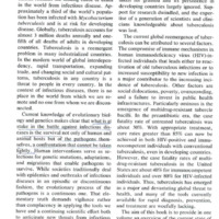 RF-TB-1.1.pdf
Position: 1540 (13 views)